Medical Physics at CNAO M Ciocca Medical Physics

: Jan. 2011 Ø Pt treatment start (experimental")

")


")

 Uniformity: ± 5% Field size 10 x 10 cm 2")

 100 SOBP Dose Phys Uniforme SOBP")


: abdominal compression, gating, 4 D imaging")
- Slides: 22
Medical Physics at CNAO M. Ciocca Medical Physics Unit, CNAO Foundation, Pavia
Ø Start of clinical commissioning (proton beams): Jan. 2011 Ø Pt treatment start (experimental phase): Sept. 2011 (protons), Nov. 2012 (carbon ions) Ø N. of pts treated: 200 (3 treat. rooms) Ø CE mark (medical device): Jul. (1 st proton protocol) and Dec. 2013 (23 clinical protocols)
Physical characterization of CNAO particle beams • Commissioning of the TPS (lat. integrated DDDs, transversal profiles, HU LUTs, simple/complex plan verification) • Determination of absorbed dose to water under reference conditions and calibration of beam monitor chambers • Determination of procedures and reference values for periodic QA checks • Dosimetry for in-vitro (cell lines) and in-vivo (mice) RB experiments
Commissioning of the TPS (Syngo RT planning, CE-marked, also used at HIT)
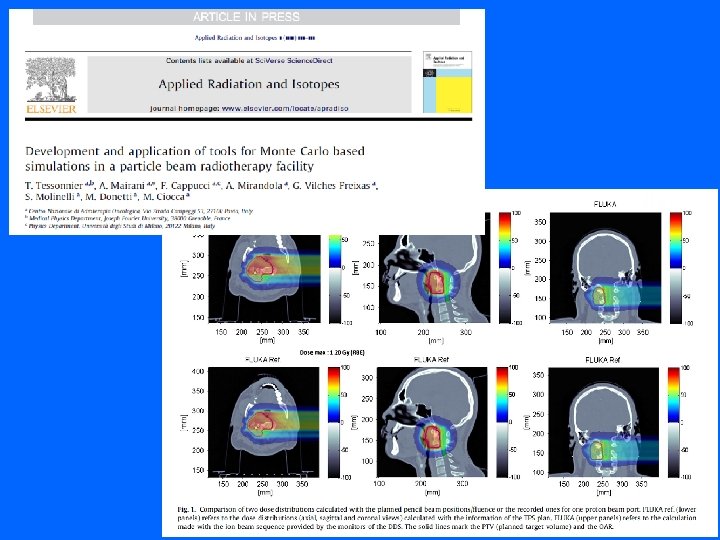
Commissioning of the TPS Physics basic beam data acquired: • experimentally • Monte Carlo simulation (FLUKA code, CNAO-HIT agreement) Experimental data Integral Depth Dose Distributions (mono-en. pencil beams) 121 Me. V/u Peakfinder water column
Ripple filter 250 Me. V/u, Ri. Fi 0 vs 3 vs 4 mm 115 Me. V/u, Ri. Fi 0 vs 4 mm
Transversal dose profiles in air
• • EBT 3 radiochromic films self-developing and water resistant dose range : 0. 01 – 10 Gy high spatial resolution (0. 2 mm) no room light sensitivity Proton beams
Carb. ion beams
Transversal dose profiles in the water phantom Pin-point IC (PTW 31014, 2 -mm diameter) E = 117. 54 Me. V/u (100 mm BP)
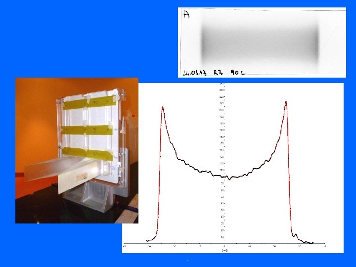
Scanned beam uniformity tests 3 -4 mm step used in the clinical practice EBT 3 films
Scanned beams (carb. ) Uniformity: ± 5% Field size 10 x 10 cm 2 Step 2 mm (1/3 FWHM)
Dose to water under ref. conditions Based on IAEA TRS-398, 2000 + formalism Hartmann GH et al. (GSI, 1999) • • Farmer-type IC, Co-60 calibrated At the isocentre, in the plateau region (2 cm), in water phantom Mono-energetic beams, different energies, 6 x 6 cm 2 homogeneous field Then, at middle SOBP (homogeneous cubic volumes) calculated by TPS
Dosimetry for RB experiments SOBP 120 dose (%) 100 SOBP Dose Phys Uniforme SOBP Dose Bio Jap Uniforme 80 60 40 20 0 0 100 200 prof. (mm H 2 O) 300
Spot position accuracy and size checks Daily QA EBT 3 films Beam energy constancy check
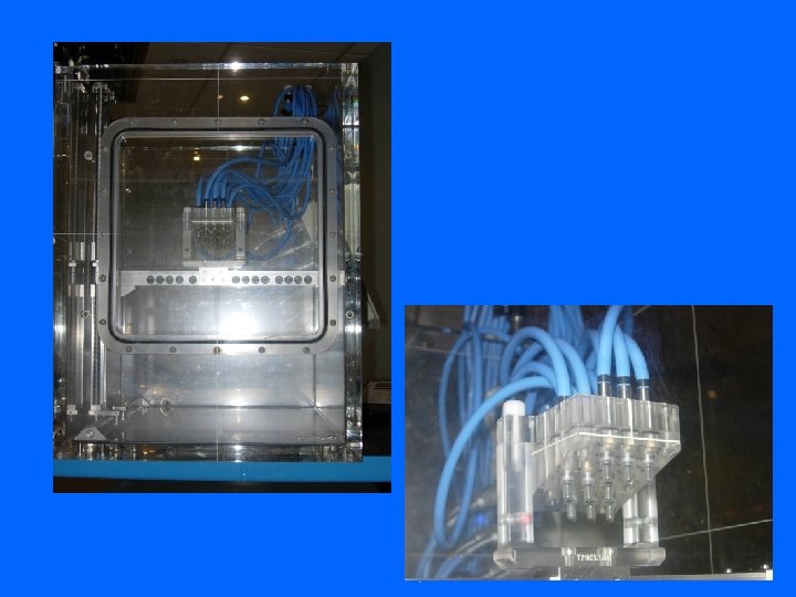
Plastic scintillator for 2 -D dosimetry
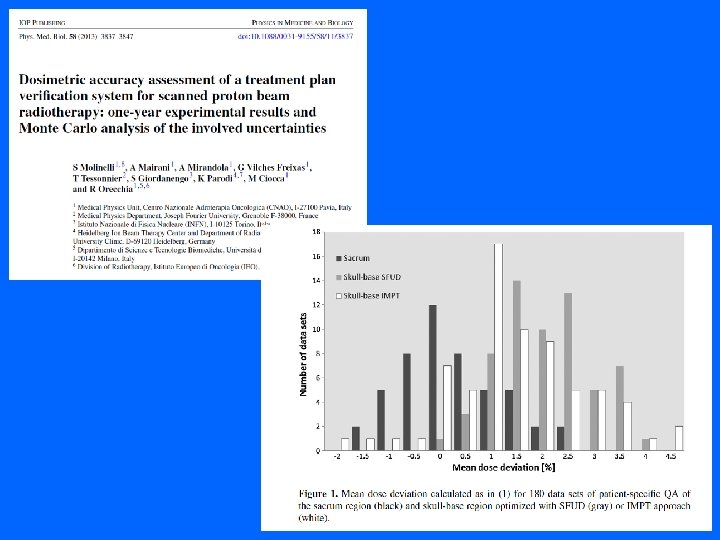
Pt-specific pre-treatment QA TPS verification plan
Future developments • Organ motion management (moving targets): abdominal compression, gating, 4 D imaging and planning • Ocular treatments • Daily patient treatment throughput optimization • Characterization of new detectors for scanned particle beams