KULTUR KALUS Pengertian kalus Sekumpulan massa sel yang



- Slides: 14
KULTUR KALUS
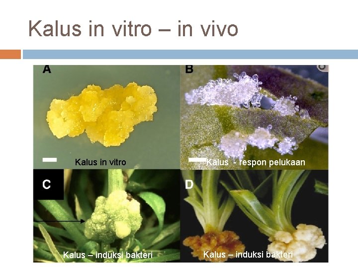
Pengertian kalus Sekumpulan massa sel yang aktif membelah dan tidak terorganisir jaringan amorf Terbentuk sebagai respon pelukaan Pada tanaman utuh : kalus adalah jaringan penutup luka fitoaleksin Bagaimana pada kultur in vitro?
Persiapan Kultur Kalus Asal eksplan: jaringan in vitro dan ex vitro hipokotil, kotiledon, daun, petiol, nodus dll. Media padat + kombinasi ZPT auksin / sitokinin rasio 1: 1 Eksplan diberi pelukaan
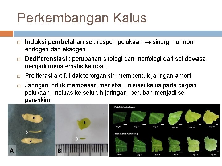
Perkembangan Kalus Induksi pembelahan sel: respon pelukaan sinergi hormon endogen dan eksogen Dediferensiasi : perubahan sitologi dan morfologi dari sel dewasa menjadi meristematis kembali. Proliferasi aktif, tidak terorganisir, membentuk jaringan amorf Jaringan induk membesar, menebal. Inisiasi kalus pada bagian pelukaan, meluas ke seluruh jaringan, berubah menjadi sel parenkim
Kalus in vitro – in vivo Kalus in vitro Kalus – induksi bakteri Kalus - respon pelukaan Kalus – induksi bakteri
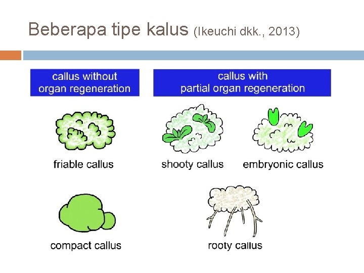
Beberapa tipe kalus (Ikeuchi dkk. , 2013)
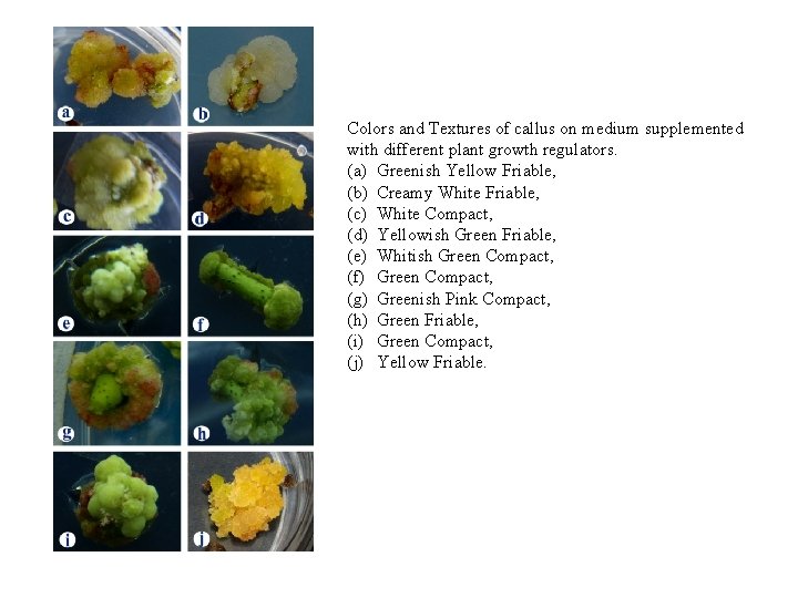
Colors and Textures of callus on medium supplemented with different plant growth regulators. (a) Greenish Yellow Friable, (b) Creamy White Friable, (c) White Compact, (d) Yellowish Green Friable, (e) Whitish Green Compact, (f) Green Compact, (g) Greenish Pink Compact, (h) Green Friable, (i) Green Compact, (j) Yellow Friable.
Pertumbuhan & pemeliharaan kalus Persentase total eksplan membentuk kalus Persentase bagian individu jaringan induk yang membentuk kalus Kecepatan pembentukan kalus Menimbang berat basah (BB) setiap periode tertentu kurva pertumbuhan analisis lanjut Menimbang berat (BK) analisis lanjut Pemeliharaan : sub kultur berdasar kurva pertumbuhan
Pertumbuhan kalus
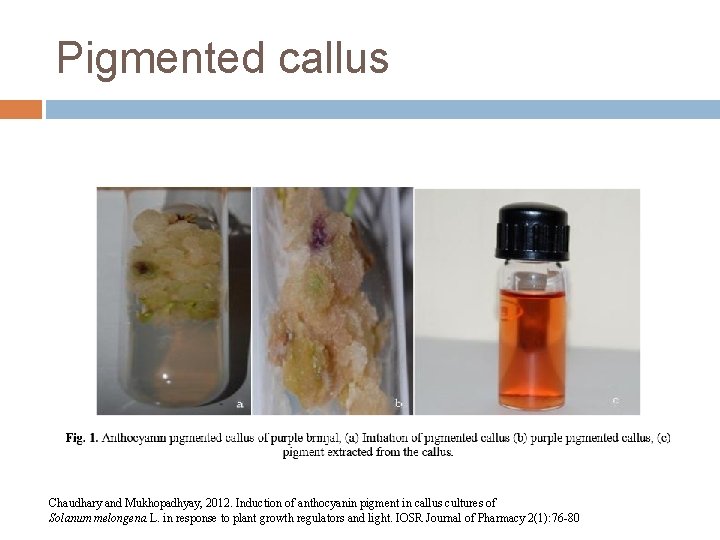
Pigmented callus Chaudhary and Mukhopadhyay, 2012. Induction of anthocyanin pigment in callus cultures of Solanum melongena L. in response to plant growth regulators and light. IOSR Journal of Pharmacy 2(1): 76 -80
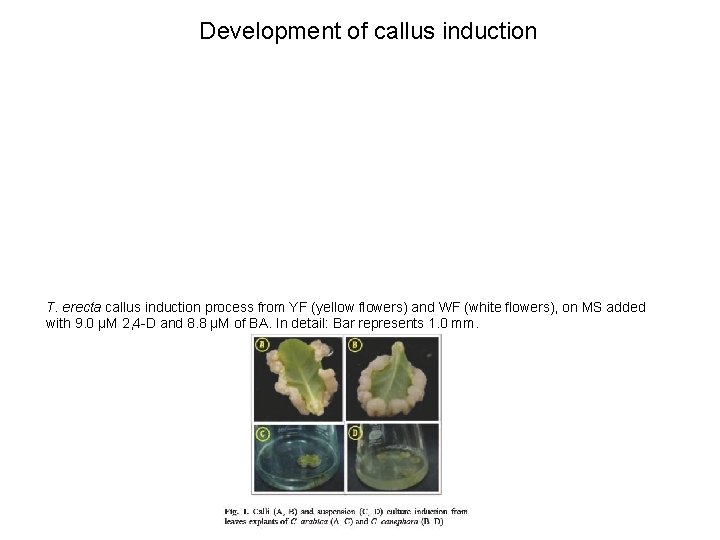
Development of callus induction T. erecta callus induction process from YF (yellow flowers) and WF (white flowers), on MS added with 9. 0 μM 2, 4 -D and 8. 8 μM of BA. In detail: Bar represents 1. 0 mm.
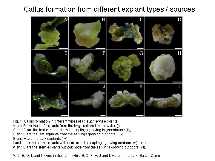
Callus formation from different explant types / sources Fig. 1. Callus formation in different types of P. euphratica explants. A and B are the leaf explants from the twigs cultured in tap water (I); C and D are the leaf explants from the saplings growing in greenhouse (II); E and F are the leaf explants from the saplings growing outdoors (III); G and H are the bark explants (IV); I and J are the stem explants with node from the saplings growing outdoors (V); and K and L are the stem explants without node from the saplings growing outdoors (VI). A, C, E, G, I, and K were in the light ; while B, D, F, H, J and L were in the dark; Bars = 2 mm.
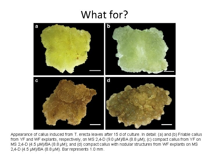
What for? Appearance of callus induced from T. erecta leaves after 15 d of culture. In detail: (a) and (b) Friable callus from YF and WF explants, respectively, on MS 2, 4 -D (9. 0 μM)/BA (8. 8 μM); (c) compact callus from YF on MS 2, 4 -D (4. 5 μM)/BA (8. 8 μM); and (d) compact callus with nodular structures from WF explants on MS 2, 4 -D (4. 5 μM)/BA (8. 8 μM). Bar represents 1. 0 mm.
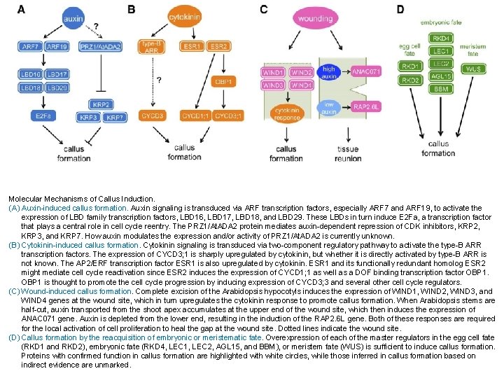
Molecular Mechanisms of Callus Induction. (A) Auxin-induced callus formation. Auxin signaling is transduced via ARF transcription factors, especially ARF 7 and ARF 19, to activate the expression of LBD family transcription factors, LBD 16, LBD 17, LBD 18, and LBD 29. These LBDs in turn induce E 2 Fa, a transcription factor that plays a central role in cell cycle reentry. The PRZ 1/At. ADA 2 protein mediates auxin-dependent repression of CDK inhibitors, KRP 2, KRP 3, and KRP 7. How auxin modulates the expression and/or activity of PRZ 1/At. ADA 2 is currently unknown. (B) Cytokinin-induced callus formation. Cytokinin signaling is transduced via two-component regulatory pathway to activate the type-B ARR transcription factors. The expression of CYCD 3; 1 is sharply upregulated by cytokinin, but whether it is directly activated by type-B ARR is not known. The AP 2/ERF transcription factor ESR 1 is also upregulated by cytokinin. ESR 1 and its functionally redundant homolog ESR 2 might mediate cell cycle reactivation since ESR 2 induces the expression of CYCD 1; 1 as well as a DOF binding transcription factor OBP 1 is thought to promote the cell cycle progression by inducing expression of CYCD 3; 3 and several other cell cycle regulators. (C) Wound-induced callus formation. Complete excision of the Arabidopsis hypocotyls induces the expression of WIND 1, WIND 2, WIND 3, and WIND 4 genes at the wound site, which in turn upregulates the cytokinin response to promote callus formation. When Arabidopsis stems are half-cut, auxin transported from the shoot apex accumulates at the upper end of the wound site, which then induces the expression of ANAC 071 gene. Auxin is depleted from the lower end, resulting in the induction of the RAP 2. 6 L gene. Both of these responses are required for the local activation of cell proliferation to heal the gap at the wound site. Dotted lines indicate the wound site. (D) Callus formation by the reacquisition of embryonic or meristematic fate. Overexpression of each of the master regulators in the egg cell fate (RKD 1 and RKD 2), embryonic fate (RKD 4, LEC 1, LEC 2, AGL 15, and BBM), or meristem fate (WUS) is sufficient to induce callus formation. Proteins with confirmed function in callus formation are highlighted with white circles, while those inferred in callus formation based on indirect evidence are unmarked.