KING SAUD UNIVERSITY MEDICAL CITY DEPARTMENT OF OBSTETRICS

KING SAUD UNIVERSITY MEDICAL CITY DEPARTMENT OF OBSTETRICS & GYNECOLOGY COURSE 482 OPERATIVE VAGINAL DELIVERIES AND CAESAREAN SECTION (C. S)

• Objectives ; • Definition of operative vaginal delivery and caesarian section • Indication of operative vaginal delivery and caesarian section • Conditions that must be fulfilled before the forceps and ventouse application • Different types of c/s • Complications of forceps , ventouse , and c/s • Trial of vaginal delivery after c/s. TOLAC • Care after operative delivery

DEFINITION It is the delivery of the fetus using an instrument through the vaginal route. Instruments could be : Forceps Vacuum Incidence of operative deliveries is 3. 5 % Indications of operative delivery MATERNAL FETAL 1. Prolonged or arrested 2 nd stage 1. Fetal distress 2. Poor maternal effort 2. Prematurity (Forceps only) 3. Maternal cardiac disease 3. Certain malpositions 4. Patients with retinal detachment or post op for similar ocular conditions
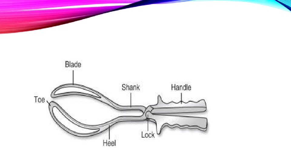
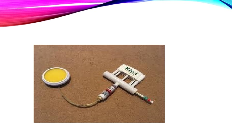
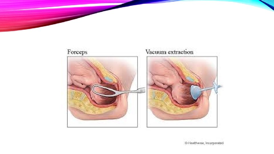

PRE-REQUISITE FORCEPS AND VENTOUSE • 1. Cervix has to be fully dilated • 2. Membranes ruptured • 3. Head has to be engaged • 4. Vertex presentation • 5. Head position known (forceps can be applied on the head for cephalic presentation or after coming head for breech presentation) Ventouse can only be applied on the head. Conditions to be fulfilled 1. Adequate analgesia 2. Empty bladder 3. Adequate episiotomy

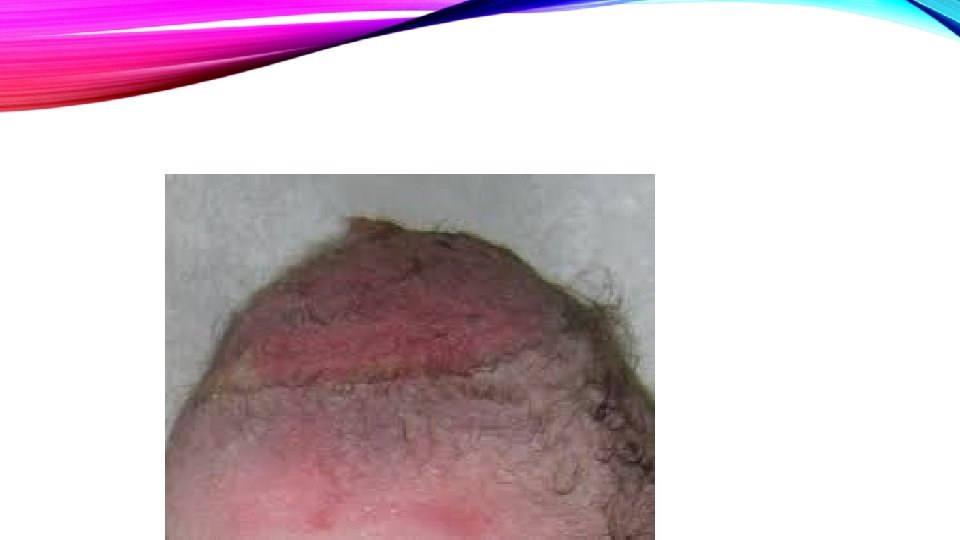
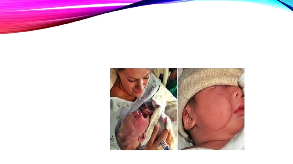
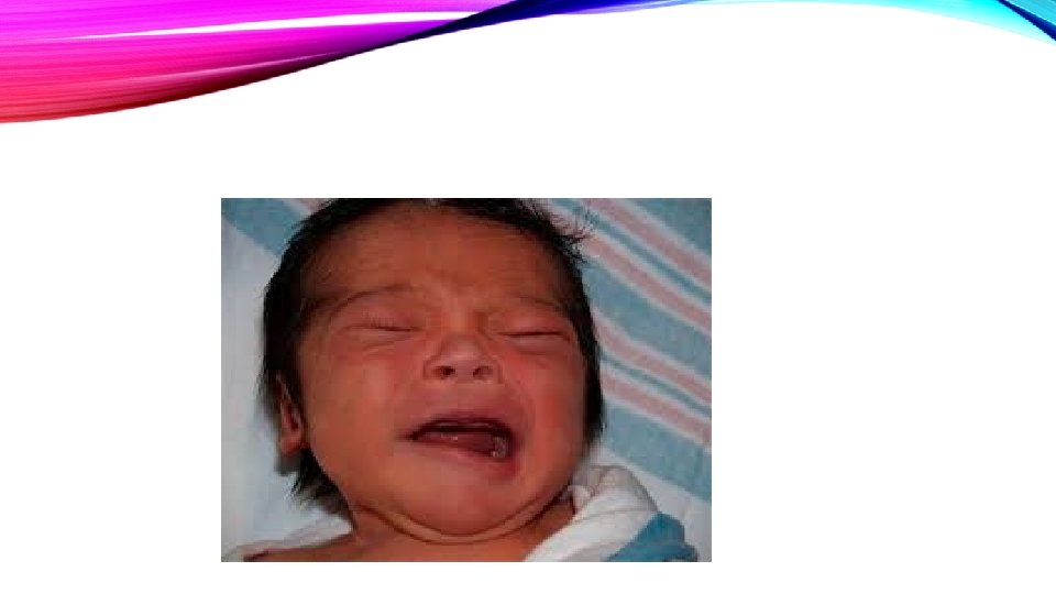
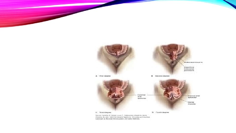
COMPLICATIONS OF INSTRUMENTAL DELIVERIES MATERNAL Genital tract lacerations, Cx, vagina, Hemorrhage FETAL Skull fractures Cephalohematoma Extensions of episiotomy Sphincter lacerations Fecal and flatus incontinence or injury to rectal mucosa Interacranial hemorrhage Facial Palsy Scalp laceration Subgalial hemorrhage

TRIAL OF INSTRUMENTAL DELIVERY Should be performed in O. R. with anesthetist present + pediatrician to resuscitate. All teams ready to proceed to C. S. in case failed instrumental delivery

CAESAREAN SECTION Rate : 25% Maternal mortality Perinatal mortality 5 – 6 per 100, 000 C/S 3/1000 USA 7/1000 U. K C. S. Could be: I. Elective C/S Planned and timed II. Emergency C/S Unplanned during labor or before the onset of labour

DIFFERENT METHODS OF PERFORMING DIFFERENT TYPES OF C/S SKIN INCISION UTERINE INCISION a. Low transverse a. Upper Segment (Classical) transverse vertical b. Midline b. Lower segment - transverse - vertical

• Indication of elective c/s • 1 -two or more previous lower segment c/s or after previous one upper segment c/s • 2 - breech presentation • 3 -placenta previa • 4 - cephalo-pelvic disproportion • 5 -triplet or higher order multiple pregnancy • 6 - infections like HIV or active genital herpes,

• Indication of emergency C/S. Non-reassuring fetal heart. Fetal distress. Failure of labor progress. Antepartum hemorrhage , abruptio placenta

COMPLICATIONS OF UPPER SEGMENT C/S 1. Bleeding 2. Organ injury Bowel Bladder Ureter 3. Adhesions formation 4. Rupture scar in future pregnancy higher than lower segment scar 5. More difficult to repair

COMPLICATIONS OF LOWER SEGMENT 1. Haemorrhage 2. Extension of incision lateral downwards 3. Organ injury bladder Bowel Ureter 4. Rupture scar 5. Abnormal placentation in future pregnancy Low lying placenta Accreta, increta, perceta 6. Adhesions specially bladder

COMMON POST OP COMPLICATIONS 1. Atelectasis 2. Infection Endometritis Wound UTI Pneumonia 3. DVT & PE

WHEN CAN A TRIAL OF LABOUR BE OFFERED AFTER C. S. 1. VBAC can be offered for non recurrent indications e. g. , fetal distress, cord prolapse, placental abruption, breech presentation. 2. Pelvic adequacy is confirmed by proper clinical methods as needed. 3. Lower Segment scar 4. Placental localization 5. Scar integrity is assured by taking proper post op history 6. Standard of care is to offer VBAC after one previous C/S and not multiple 7. Safe set up: Tertiary care center which can perform emergency C. S as needed. 8. Patients approval

MEASURES TO REDUCE C. S. RATE Proper antenatal care For early detection and management of conditions that lead to C. S. rate e. g. controlling macrosomia in diabetes early detection of HTN. Post term ect. Performing ECV for breeches. Prevent infections: Prophylactic Ab + Aseptic technique Prevention of anemia To prevent DVT. : TEDS stocking Thromboprophylaxis

POST CARE 1. VS hourly x 4 hours 2. I. V. fluids 3. Analgesia 4. Checking Fundus + Lochia 5. Urine output + catheter care 6. Wound care 7. Early ambulation 8. Antibiotics 9. Thromboprohylaxis 10. Breast care and breast feeding
- Slides: 23