Golgi Apparatus The post office of the cell


 • Doctor")


- Slides: 15
Golgi Apparatus “The post office of the cell” Tamboli Alija Z. Dept of Zoology, S. M. Joshi Collge , Hadapsar. F. Y. B. Sc
Camillo golgi
Camillo Golgi- discoverer of golgi apparatus (july 7, 1843 -jan 21, 1926) • Doctor Camillo golgi on july 15, 1898 discovered the golgi apparatus or golgi complex. • He got a noble prize in physiology and medicine in 1906. • Golgi discovered the method of staining nervous tissue that would stain limited number of cells at random. • This enabled him to view the paths of nervous cells for the first time. • He called his discovery the black reaction which later received the name golgi method or golgi stain or silver staining technique. • Golgi also discovered tendon sensory organ that bears his name golgi receptor • Using his staining technique golgi identified the intracellular reticular apparatus in 1898 which bears his name golgi apparatus.
Golgi Complex and its discovery…. • The golgi apparatus or golgi complex is found in most of the cells. • It is the another packing organelle like the endoplasmic reticulum. • It was named after the scientist who discovered it. • Golgi in 1898 in the nerve cells of barn owl and cat by metallic impregnation method. • Many scientist thought it to be artefact produced by the staining technique. • The job of golgi apparatus is to process and bundle macromolecule like proteins and lipid as they are synsthesized within the cells. • The golgi apparatus is sometimes are called as post office of the cell since one major function is to modify, sort, and package proteins to be secreted.
Structure of golgi body…
Structure… • Golgi bodies varies in size and form in different cell types, but usually has similar organization for kind of cells. For example it is well developed in secretory and nerve cells but is rather small in muscle cells. • Electron microscope show it as a central stack of flattened sacs or cisternae and many peripheral tubes and vesicles.
Cisternae… • The cisternae vary in number from 3 -7 in most animal cells and from 10 -24 in plant cells. • They are equally spaced in the pile so that they are nearly parallel to one another , having 200 -300 angstrom. • In certain cells, the intercisternial spaces contain a layer of parallel fibres called intercisternial elements. • These support cisternae and maintain spacing between them. • The cisternae may be flat but sometimes curved having a distinct polarity with a convex face towards the cell membrane and a concave face towards the nucleus. • They are free of ribosomes and have swollen ends. • They look like the SER and continuous at with it at certain places • This suggests golgi body is derieved from SER.
Tubules and vesicles…. • Tubules are shprt, anastomosing tubules arise from thhe periphery of the cisternae. Some of these enlarge to form vesicles. • Vesicles lie near the ends and concave surface of golgi complex. • They are pinched of from the tubules of the cisternae and are of two types: • Smooth vesicles- they have smooth surface and contain secretion of the cell. They are also called secretory vesicles. They arise from the end of cisternal tubules. • Coated vesicles- they have rough surface. They also arise from cisternal tubules their function is not clear.
Vesicles… • The membrane surrounds an area of fluid where the complex molecules i. e. proteins , sugars , enzymes are stored and changed. • Because the golgi complex absorbs vesicles from the rough endoplasmic reticulum, and ribosomes are also find ribosomes in those pancakes stacks.
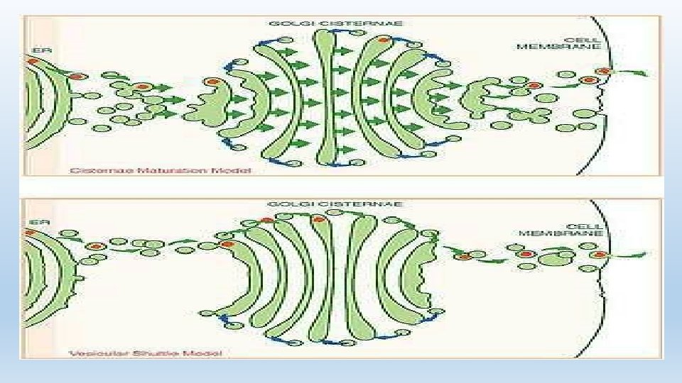
Working with Rough ER…
Working with Rough ER… • The Golgi complex works closely with the rough endoplasmic reticulum. • When a protein is made in ER, something called a transition vesicle is made. • The vesicle or sac floats through the cytoplasm to the golgi apparatus and is absorbed. • After the golgi does it work on the molecules inside the sac , a secretory vesicle is created and released into the cytoplasm. • From there the vesicle moves to thhe cell membrane and the molecules are released out of the cell.
Functions of golgi apparatus…. • Formation of secretory vesicles. • Synthesis of carbohydrates. • Formation of glycoproteins. • Formation of lipoproteins. • Addition to cell membrane. • Membrane transformation. • Formation of cell wall. • Formation of lysosomes. • Acrosome formation.
Functions… • formation of yolk and cortical granules. • Formation of nematocysts and trichocyts. • Storage of secretions. • Absorption of materials. • Location of enzymes
Thanks!