FRACTURE HEALING Dr Sowmya Uthaiah Assistant professor Dept


- Slides: 17
FRACTURE HEALING Dr. Sowmya Uthaiah Assistant professor Dept of pathology Yenepoya medical college
Fractures • Definition : A fracture is defined as loss of bone integrity due to mechanical injury and/or diminished bone strength
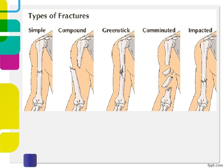
Types of fratures 1. Simple: the overlying skin is intact 2. Compound: the bone communicates with the skin surface 3. Comminuted: the bone is fragmented 4. Displaced: the ends of the bone at the fracture site are not aligned
Types of fractures……… 5. Stress: a slowly developing fracture that follows a period of increased physical activity in which the bone is subjected to repetitive loads 6. “Greenstick”: extending only partially through the bone, common in infants when bones are soft 7. Pathologic: involving bone weakened by an underlying disease process, such as a tumor
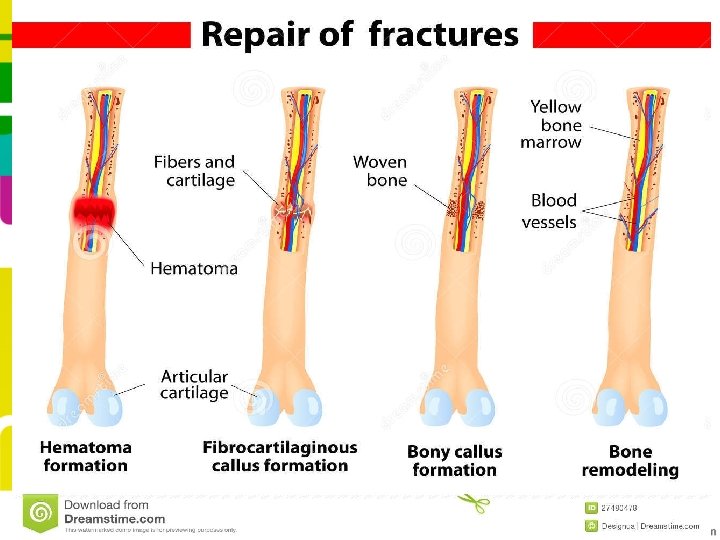
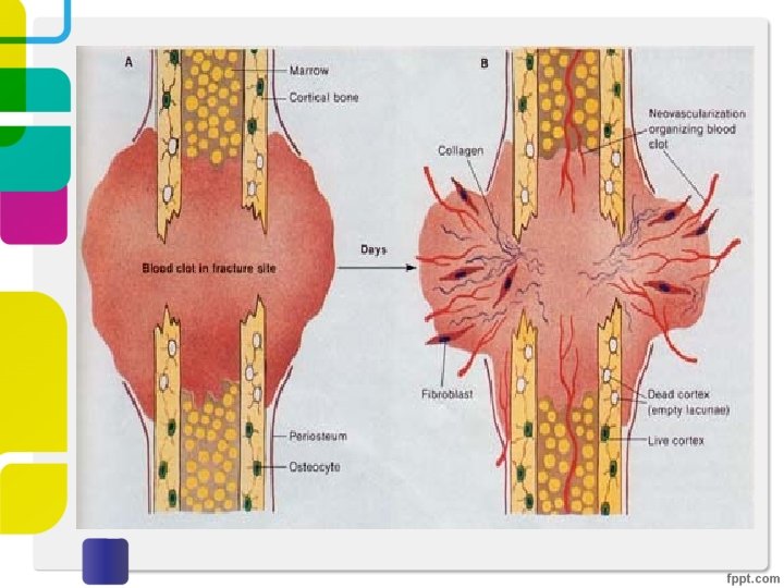
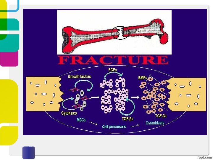
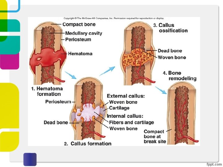
HEALING OF FRACTURES • Immediately after fracture----rupture of blood vessels------hematoma----- fills the fracture gap and surrounds the area of bone injury. • clotted blood provides ü a fibrin mesh ü sealing off the fracture site ü creates a framework for the influx of inflammatory cells ingrowth of fibroblasts and new capillaries
degranulated platelets and migrating inflammatory cells release PDGF, TGF-β, FGF, and other factors activate osteoprogenitor cells in the periosteum, medullary cavity, and surrounding soft tissues stimulate osteoclastic and osteoblastic activity
By the end of the first week -the major changes are ü organization of the hematoma ü matrix production in adjacent tissues ü remodeling of the fractured ends of the bone fusiform and predominantly uncalcified tissue called soft tissue callus or procallus ü Provides some anchorage between the ends of the fractured bones ü No structural rigidity for weight bearing
After approximately 2 weeks the soft tissue callus is transformed into a bony callus § activated osteoprogenitor cells deposit subperiosteal trabeculae of woven bone oriented perpendicular to the cortical axis and within the medullary cavity § activated mesenchymal cells in the soft tissues and bone surrounding the fracture line- differentiate into chondrocytes- make fibrocartilage and hyaline cartilage. Bony callus reaches its maximal girth at the end of the second or third week and helps to stabilize the fracture site
• The newly formed cartilage along the fracture line undergoes enchondral ossification contiguous network of bone with newly deposited bone trabeculae in the medulla and beneath the periosteum ü Fractured ends are bridged ü Mineralizes – stiffness and strength of the callus increases ü Controlled weight bearing is tolerated
Bone remodelling • early stages of callus formation, an excess of fibrous tissue, cartilage, and woven bone is produced • callus matures and is subjected to weight-bearing forces----portions that are not physically stressed are resorbed-----callus is reduced in size • the shape and outline of the fractured bone are reestablished as lamellar bone • restoration of the medullary cavity
Defects in healing • Displaced and comminuted fractures- deformity • Delayed union or nonunion- Inadequate immobilization permits movement of the callus---prevents its normal formation. • False joint or pseudoarthrosis-nonunion persists---malformed callus----cystic degeneration----luminal surface lined by synovial-like cells • infection of the fracture site esp open fractures • Malnutrition and skeletal dysplasia, osteoporosis and osteomalacia - hinder fracture healing