Evaluation of Knee Injuries Dr Alan A Zakaria




- Slides: 32
Evaluation of Knee Injuries Dr. Alan A. Zakaria, D. O. , M. S. 1080 Kirts Blvd. , Suite 400 Troy, Mi. , 48084 Team Physician United States Soccer Federation University of Michigan Men’s and Women’s Soccer
Objective Identify main anatomic components of the knee Perform basic knee exam along with special tests Identify common knee injury patterns and their physical exam findings.
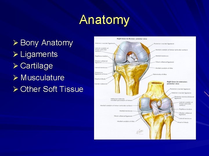
Anatomy Ø Bony Anatomy Ø Ligaments Ø Cartilage Ø Musculature Ø Other Soft Tissue
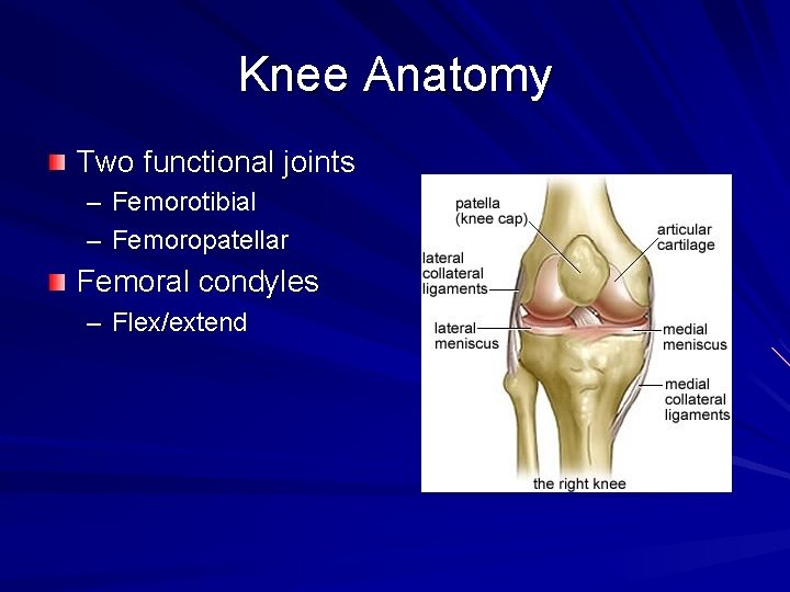
Knee Anatomy Two functional joints – Femorotibial – Femoropatellar Femoral condyles – Flex/extend
Knee Anatomy Patella – Sesamoid with two concave surfaces and vertical ridge – Increases efficiency of extension
Knee Anatomy: Anterior Cruciate Ligament (ACL) Run inferior, anterior, and medially Arises from medial aspect lateral femoral condyle Insert lateral to medial tibial eminence Restrains anterior subluxation of tibia on femur
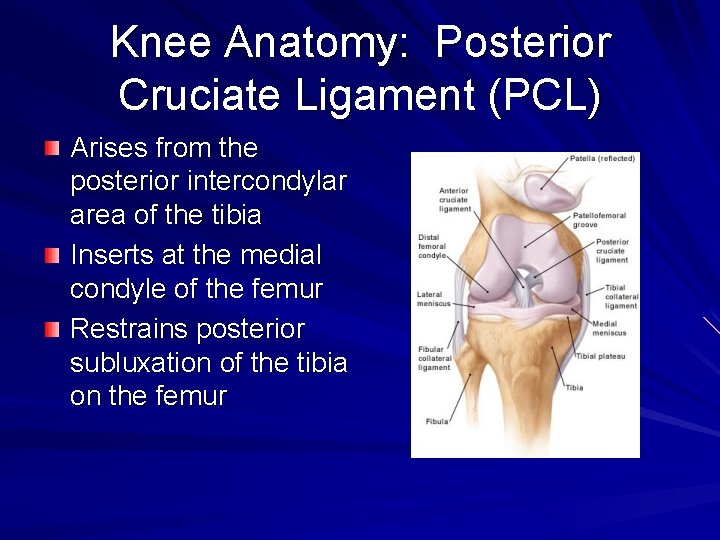
Knee Anatomy: Posterior Cruciate Ligament (PCL) Arises from the posterior intercondylar area of the tibia Inserts at the medial condyle of the femur Restrains posterior subluxation of the tibia on the femur
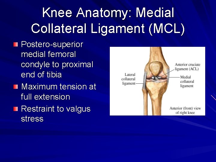
Knee Anatomy: Medial Collateral Ligament (MCL) Postero-superior medial femoral condyle to proximal end of tibia Maximum tension at full extension Restraint to valgus stress
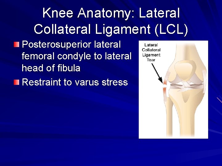
Knee Anatomy: Lateral Collateral Ligament (LCL) Posterosuperior lateral femoral condyle to lateral head of fibula Restraint to varus stress
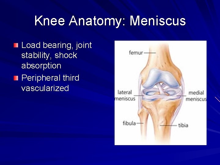
Knee Anatomy: Meniscus Load bearing, joint stability, shock absorption Peripheral third vascularized
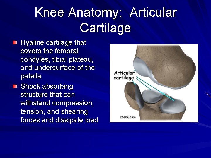
Knee Anatomy: Articular Cartilage Hyaline cartilage that covers the femoral condyles, tibial plateau, and undersurface of the patella Shock absorbing structure that can withstand compression, tension, and shearing forces and dissipate load
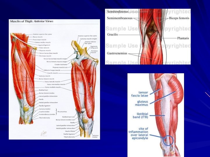
Knee Anatomy: Muscles Quadriceps Hamstrings Iliotibial tract Pes Anserine
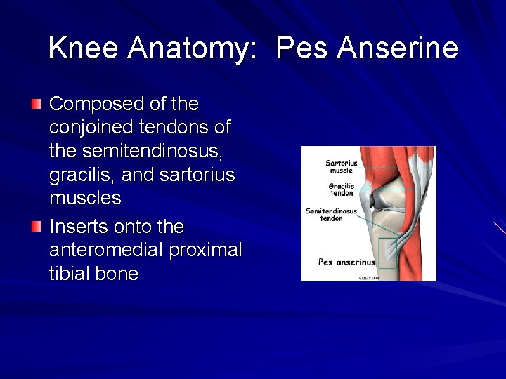
Knee Anatomy: Pes Anserine Composed of the conjoined tendons of the semitendinosus, gracilis, and sartorius muscles Inserts onto the anteromedial proximal tibial bone
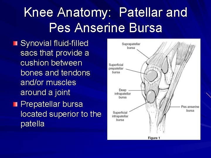
Knee Anatomy: Patellar and Pes Anserine Bursa Synovial fluid-filled sacs that provide a cushion between bones and tendons and/or muscles around a joint Prepatellar bursa located superior to the patella
Patient History. Ø Nature of symptoms Ø Age Ø Location Ø Occupation or Ø Timing/Duration recreational activities Ø Clicking or popping Ø Locking or catching Ø Giving Way Ø Swelling Ø Quality and Quantity Ø Trauma or preceding event Ø Exacerbating or relieving factors Ø Past History
Physical Exam Ø Observation Ø Range of Motion Ø Palpation Ø Strength Testing Ø Special Testing
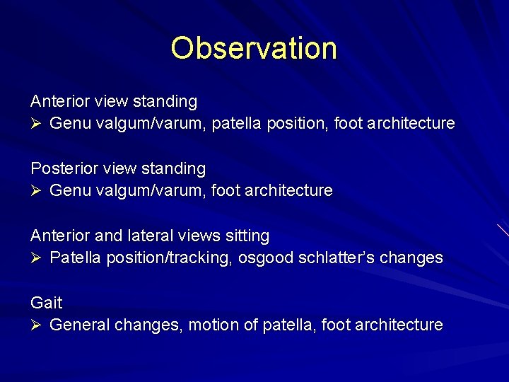
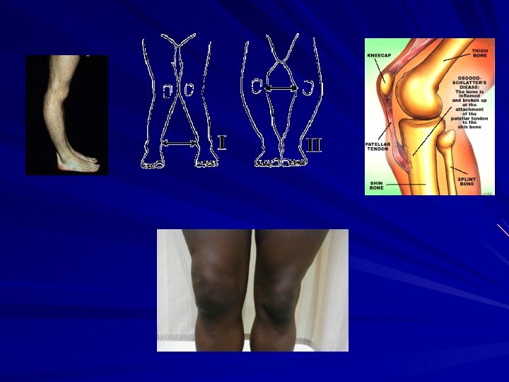
Observation Anterior view standing Ø Genu valgum/varum, patella position, foot architecture Posterior view standing Ø Genu valgum/varum, foot architecture Anterior and lateral views sitting Ø Patella position/tracking, osgood schlatter’s changes Gait Ø General changes, motion of patella, foot architecture
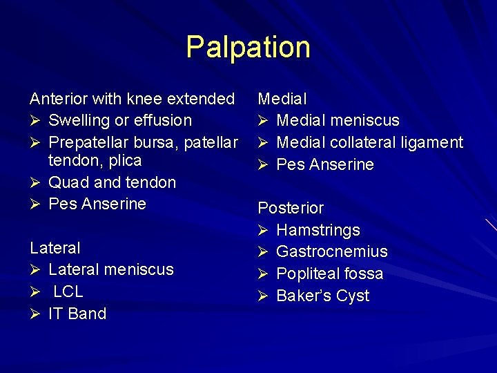
Palpation Anterior with knee extended Ø Swelling or effusion Ø Prepatellar bursa, patellar tendon, plica Ø Quad and tendon Ø Pes Anserine Lateral Ø Lateral meniscus Ø LCL Ø IT Band Medial Ø Medial meniscus Ø Medial collateral ligament Ø Pes Anserine Posterior Ø Hamstrings Ø Gastrocnemius Ø Popliteal fossa Ø Baker’s Cyst
Range of Motion Ø Flexion – 135 -140 degrees Ø Extension – 0 degrees
Strength Testing (5 point scale) Flexion Ø Hamstring Ø Gracilis, sartorius Ø Popliteus Ø Gastrocnemius Extension Ø Quadriceps Ø Tensor Fasciae Latae
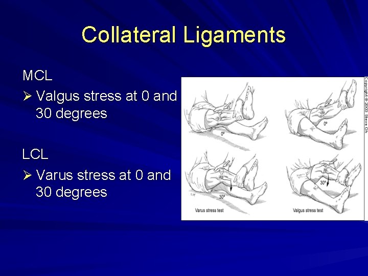
Collateral Ligaments MCL Ø Valgus stress at 0 and 30 degrees LCL Ø Varus stress at 0 and 30 degrees
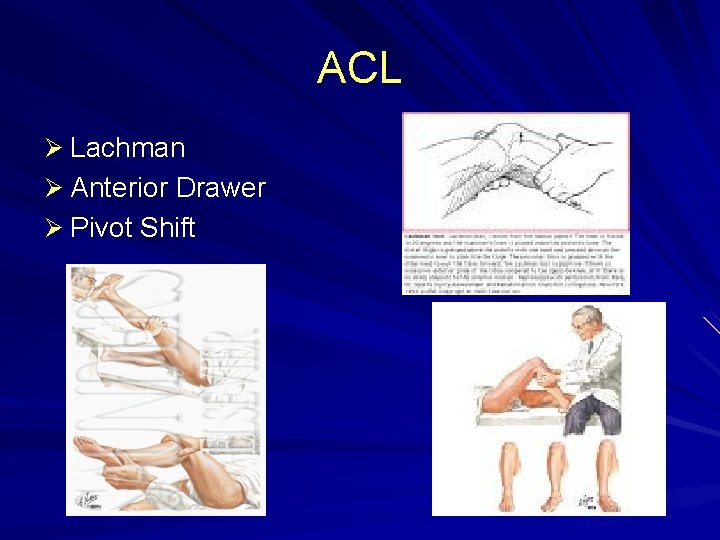
ACL Ø Lachman Ø Anterior Drawer Ø Pivot Shift
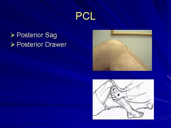
PCL Ø Posterior Sag Ø Posterior Drawer
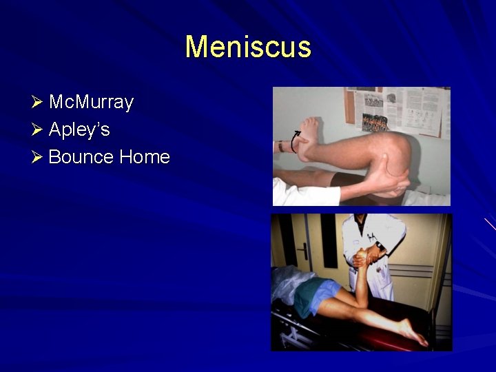
Meniscus Ø Mc. Murray Ø Apley’s Ø Bounce Home
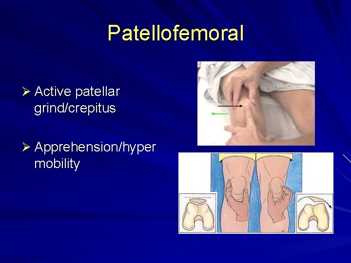
Patellofemoral Ø Active patellar grind/crepitus Ø Apprehension/hyper mobility
Others Ober’s Test
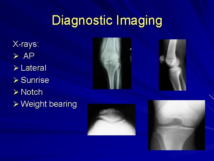
Diagnostic Imaging X-rays: Ø AP Ø Lateral Ø Sunrise Ø Notch Ø Weight bearing
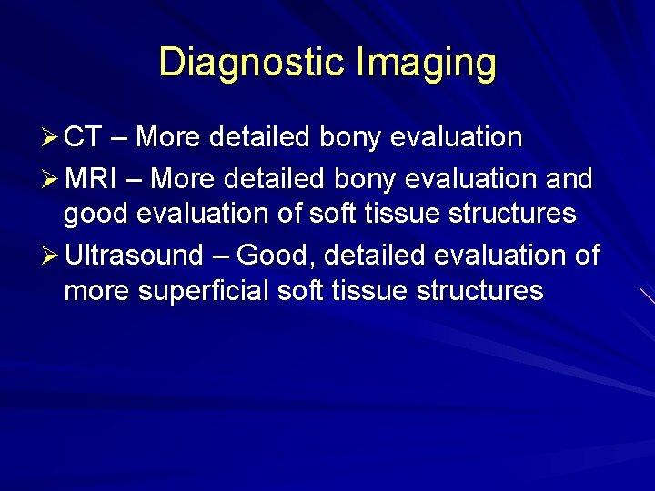
Diagnostic Imaging Ø CT – More detailed bony evaluation Ø MRI – More detailed bony evaluation and good evaluation of soft tissue structures Ø Ultrasound – Good, detailed evaluation of more superficial soft tissue structures
Summary Knee Anatomy Basic physical exam Special tests
Thank You!