Electrocardiography Dr Arun Goel Associate professor Department of

- Slides: 29
Electrocardiography Dr. Arun Goel Associate professor Department of Physiology
Specific learning objectives • Introduction • History • Normal ECG • Procedure • Limb Leads
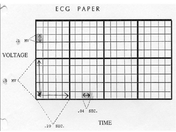
ECG, EKG It is the procedure of recording the electrical activity of the heart. The electrode combination records the difference of potential difference at two sites on the body. The potential differences are produced due to the electrical activity of the heart. Electrocardiograph is the machine Electrocardiogram is the record The characteristic shape and timing of the ECG waves are due to the spread of wave of depolarization and repolarization associated with each heart beat.
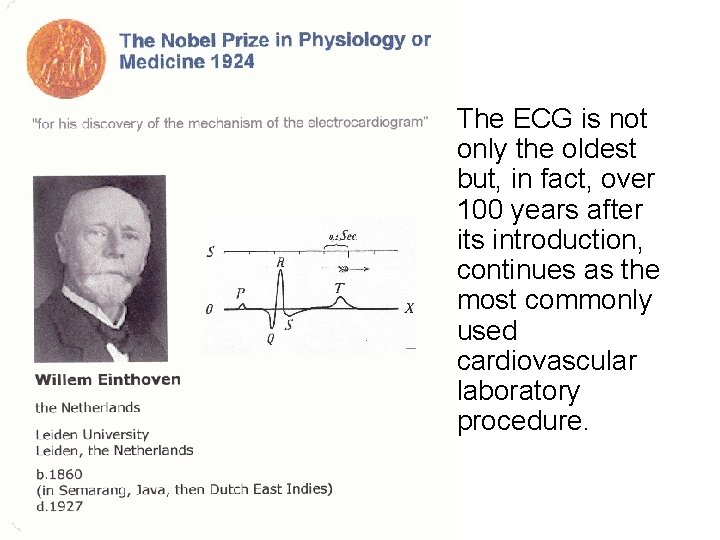
• The ECG is not only the oldest but, in fact, over 100 years after its introduction, continues as the most commonly used cardiovascular laboratory procedure.
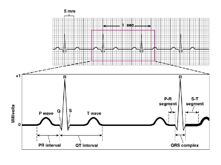
What does the ECG look like?
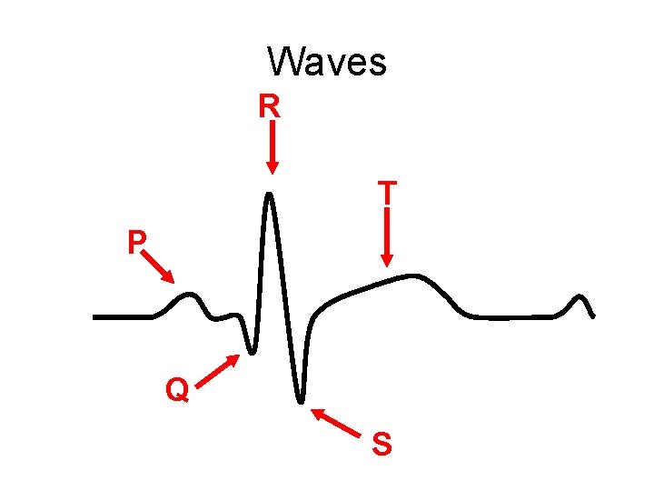
Waves R T P Q S
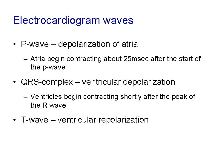
Electrocardiogram waves • P-wave – depolarization of atria – Atria begin contracting about 25 msec after the start of the p-wave • QRS-complex – ventricular depolarization – Ventricles begin contracting shortly after the peak of the R wave • T-wave – ventricular repolarization
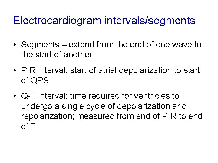
Electrocardiogram intervals/segments • Segments – extend from the end of one wave to the start of another • P-R interval: start of atrial depolarization to start of QRS • Q-T interval: time required for ventricles to undergo a single cycle of depolarization and repolarization; measured from end of P-R to end of T
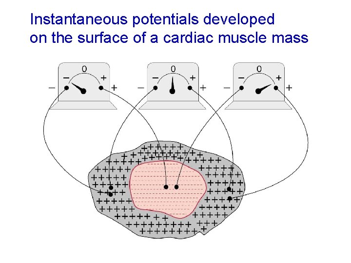
Instantaneous potentials developed on the surface of a cardiac muscle mass
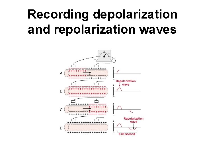
Recording depolarization and repolarization waves
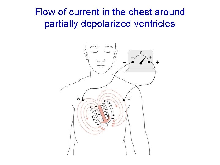
Flow of current in the chest around partially depolarized ventricles
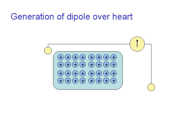
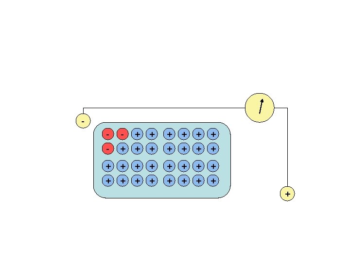
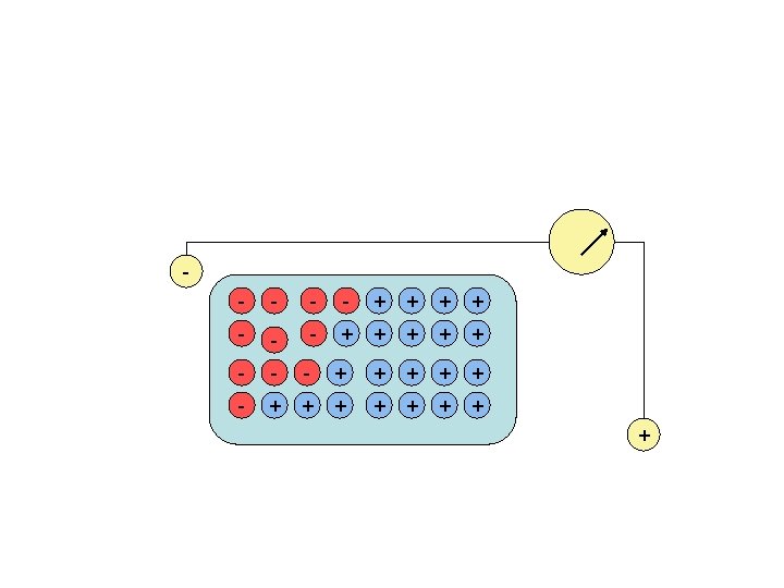
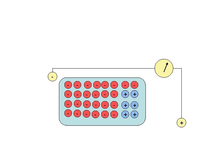
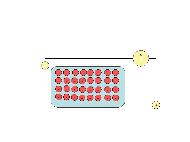
Generation of dipole over heart + + + + + + + +
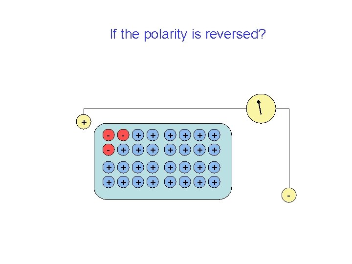
If the polarity is reversed? + - - + + + + + + + + -
Remember The representation of the electrical activity of the heart is critically dependent on the position of the electrodes. The same electrical activity will look different on different electrode combinations A combination of electrodes for recording is called a LEAD
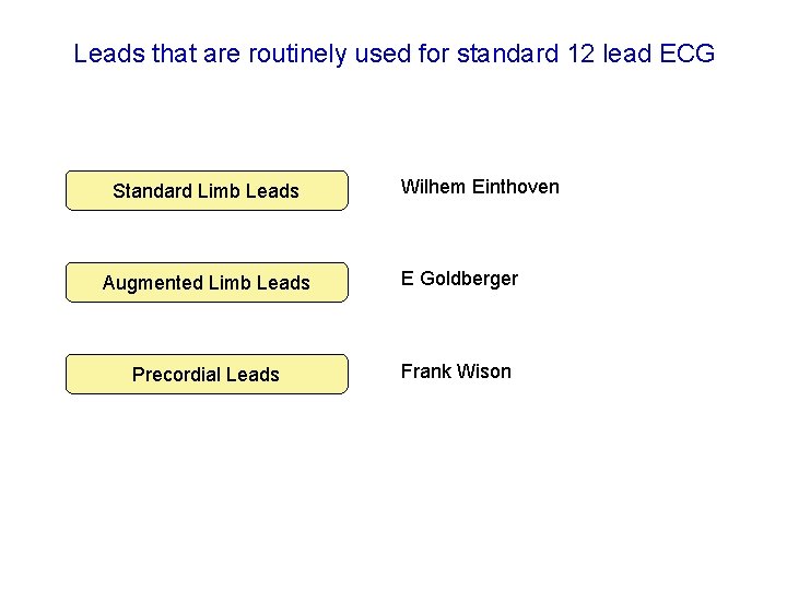
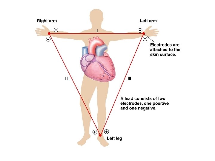
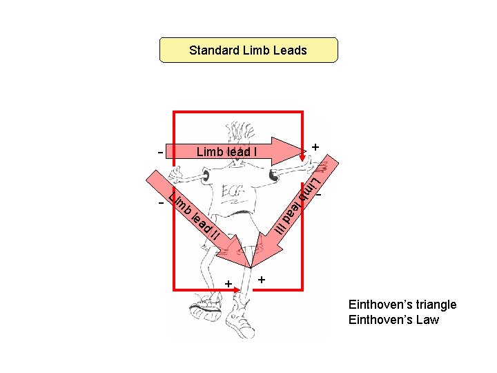
Leads that are routinely used for standard 12 lead ECG Standard Limb Leads Wilhem Einthoven Augmented Limb Leads E Goldberger Precordial Leads Frank Wison
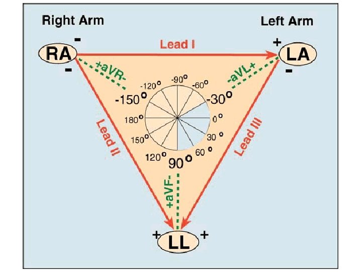
Standard Limb Leads - Lim Li b m b d ad - III le lea - + Limb lead I II + + Einthoven’s triangle Einthoven’s Law
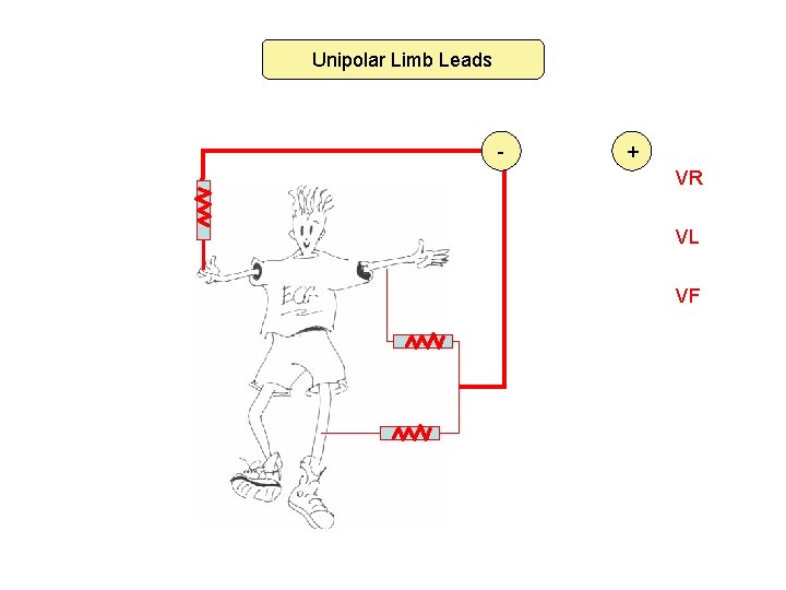
Unipolar Limb Leads - + VR VL VF
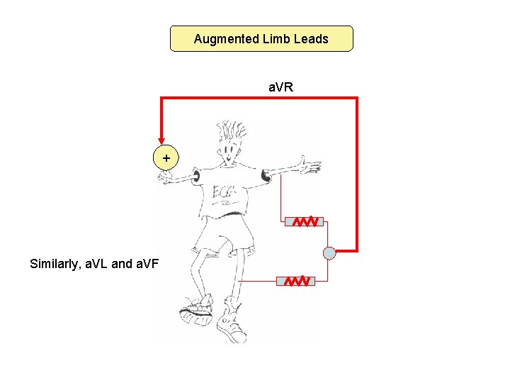
Augmented Limb Leads a. VR + Similarly, a. VL and a. VF
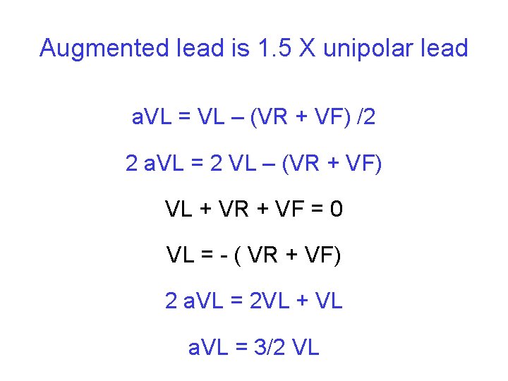
Augmented lead is 1. 5 X unipolar lead a. VL = VL – (VR + VF) /2 2 a. VL = 2 VL – (VR + VF) VL + VR + VF = 0 VL = - ( VR + VF) 2 a. VL = 2 VL + VL a. VL = 3/2 VL
Thank You