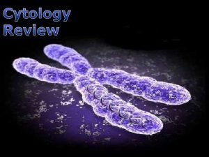
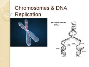
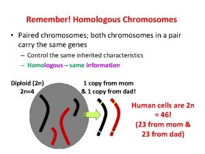
Chromosomes and chromatin Chromosomes organize and package genes


- Slides: 24

Chromosomes and chromatin

Chromosomes organize and package genes inside cells • Bind packaging proteins to DNA to make it more compact. – Histones +DNA = chromatin in eukaryotes – Virion proteins in viruses – HU (? ) or other proteins in bacteria • Loop chromatin and attach it to a matrix in nuclei

Bands and specialized regions of human chromosomes Human chromosome 11: 125 Mb, 180 c. M

Human chromosomes, ideograms Mitotic chromosomes are spread and stained with Geimsa. Those that stain are shown in black. G-bands (more A+T rich).

Human chromosomes, spectral karyotype Reagents specific to each chromosome. Chromosome painting.

Identifying translocations http: //www. ncbi. nlm. nih. gov/disease/

Distinctive and common features of chromosomes • Distinctive proteins and DNA sequences have been used to develop chromosome painting reagents. • Genomic DNA in vertebrates has long (megabase) stretches of G+C rich DNA, and other long stretches of A+T rich DNA – Called isochores • Virtually all this DNA is organized into chromatin, which has a common fundamental structure.

Chromatin Structure

Principal proteins in chromatin are histones H 3 and H 4 : Arg rich, mostly conserved sequence H 2 A and H 2 B : Slightly Lys rich, fairly conserved H 1 : very Lys rich, most variable in sequence between species

Histone structure and function

Histone interactions via the histone fold

Nucleosomes are the subunits of the chromatin fiber • Experimental evidence: – Beads on a string in EM – Micrococcal nuclease digestion

General model for the nucleosomal core

A string of nucleosomes

Detailed structure of the nucleosomal core

Higher order chromatin structure Histone H 1 associates with the linker DNA, and may play a role in forming higher order structures.

Alterations to chromatin structure are key steps in regulation

Phosphorylation of histones

Acetylation and Deacetylation of lysines in proteins

Acetylation and Deacetylation of histones

Effects of histone modifications • Highly acetylated histones are associated with actively transcribed chromatin – Acetylation of histone N-terminal tails may affect the ability of nucleosomes to associate in higher -order structures – The acetylated chromatin appears to be more “open”, and accessible to transcription factors and polymerases – HATs are implicated as co-activators of genes in chromatin, and HDACs are implicated as corepressors

Matrix and scaffold Mitotic chromosomes, with some DNA released In interphase chromosomes, at least some DNA is attached to a matrix

Chromosome localization in interphase In interphase, chromosomes appear to be localized to a sub-region of the nucleus.

Gene activation and location in the nucleus • Condensed chromatin tends to localize close to the centromeres – Pericentromeric heterochromatin • Movement of genes during activation and silencing – High resolution in situ hybridization – Active genes found away from pericentromeric heterochromatin – Silenced genes found associated with pericentromeric heterochromatin
 Linked genes and unlinked genes
 Linked genes and unlinked genes
 Glomerulus
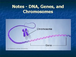
 The relationship between genes dna and chromosomes
 Chromosomes genes and basic genetics foldable answer key
 Dna, genes and chromosomes relationship
 What is the relationship between dna chromosomes and genes
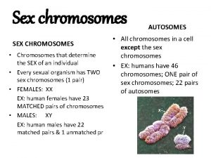
 Genes located on the sex chromosomes
 Genes chromosome
 Dna chromosomes genes diagram
 Sex nn
 Chromatin vs chromozom
 Chromatin vs chromozom
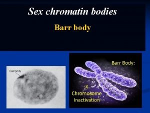
 Barrovo telisko
 Chromatin body
 Chromatin in a sentence
 Malaria parasite in thick film
 Chromatin
 Parasites
 Chromatin draws together to create
 Chromatin
 Phases of mitosis
 Chromatin states
 Chromatin immunoprecipitation
 반보존적 복제

