Cell signaling Color Index Important Extra Information Doctors


 1 - The ligand ( signal or primary messenger )")

 Same Cell but, Different Pathways (continued)")
 GTP-Dependant Regulatory Proteins - come in between the receptor & 2 nd messenger.")


")

")


, Acetylecholine")


- Slides: 24
Cell signaling – Color Index: § Important. § Extra Information. § Doctors slides. 436 Biochemistry team
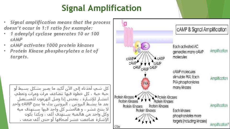
Objectives: • • • Differentiate different steps in signaling pathways Describe the second messenger systems Recognize the function of signaling pathways for • Signal transmission Amplification • Discuss the role of signaling pathways in regulation and integration of metabolism •
NO CELL LIVES IN ISOLATION* • Cells communicate with each other. • Cells send & receive information (signals)** • Information is relayed (received) within cell to produce a response. Recognition of signal* By receptors Transduction Change of external signal into intracellular message with amplification & formation of second messenger** General concept Cell signaling is part of a complex system of communication that governs basic activities of cells and coordinates cell actions. The ability of cells to understand correctly respond to their microenvironment is the basis of development, tissue repair, and immunity as well as normal tissue homeostasis. Notes: *Because cell survival depends on communication. **Dr’s note: If cells are adjacent to each other, they can communicate without signals Signalling Process # ﻟﻠﺮﺑﻂ Transduction= transformation= change Effect Modification of cell metabolism & function*** Dr’s notes : *Signals which are lipid soluble can go directly to the cell without the recognition by a receptors. (No recognition step) **The first messenger is called (Signals) while the second messenger is called (intracellular signals) ***The modification of cell metabolism & function is done by the second messenger (Intracellular signals)
General signaling pathway (cascade) 1 - The ligand ( signal or primary messenger ) will bind to the receptor 2 - the receptor will recognize this signal. 3 - This binding will stimulates the transduction by the second messenger, and this transduction includes: • Cellular responses • Changes in gene expression. Biochemical cascade: a series of biochemical reactions, in which a product of the previous step is the substrate of the next. 1 2 3 Recognition More in Next slide Performed by receptors Ligand (hormone or neurotransmitter) will produce response only in cells that have receptors for this particular ligand. Each cell has a specific set of receptors 4
More in next slide
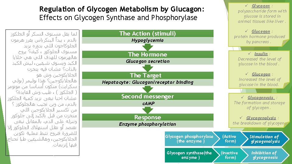
Different Responses to the Same Signaling Molecule: (B) Same Cell but, Different Pathways (continued) Its secretion is stimulated by: Hypoglycemia (deficiency of glucose in the bloodstream) Glucagon (hormone) secretion 1 Hepatocyte (liver cells) : They have glucagon receptor 2 Second messenger: c. AMP ( cyclic Adenosine Monophosphate ) Response: Enzyme phosphorylation (add P group) P P Glycogen synthase 3 (Inactive form) Inhibition of glycogenesis Formation of glycogen Both provide more glucose to blood Glycogen phosphorylase 4 (Active form) Stimulation of glycogenolysis breakdown of glycogen to glucose 1. Glucagon is a peptide hormone, produced by alpha cells of the pancreas. The effect of glucagon is to make the liver release the glucose it has stored in its cells into the bloodstream, with the net effect of increasing blood glucose. 2. A receptor coupled with G-protein *more in next slide* 3. Is an enzyme involved in converting glucose to glycogen therefore glucagon makes it INACTIVE. 4. Glycogen phosphorylase is one of the phosphorylase enzymes. It breaks up glycogen into glucose subunits.
(G-Proteins) GTP-Dependant Regulatory Proteins - come in between the receptor & 2 nd messenger. - Regulate the formation of synthesis of 2 nd messenger G-Proteins : Trimeric ( 3 subunits α, β, γ ) membrane proteins. Types according to its function : G-stimulatory (Gs) and G-inhibitory (Gi) binds to GTP/GDP Forms of G-Proteins Inactive form Trimeric –bound GDP (αβγ/GDP) Active form ONLY α-bound GTP (α/GTP) ( β , γ separate from α) - The α-subunit has intrinsic GTPase activity ( act as an Enzyme ), resulting in hydrolysis of GTP into GDP and inactivation of G-proteins Note: GTP: Guanosine triphosphate. GTP is essential to signal transduction, in particular with G-proteins, in second-messenger mechanisms where it is converted to guanosine diphosphate (GDP) through the action of GTPases.
Two important second messenger systems: Adenylyl cyclase system. Remember Second messenger: (intracellular signal Formed during transduction) intracellular signaling molecules released within the cell in response to a hormone to trigger physiological changes. Calcium phosphatidylinositol system.
First: Adenylyl cyclase system Signal: Hormones or neurotransmitters are primary messengers § Hormones or neurotransmitters such as : Glucagon & Epinephrine § Or toxins such as : Cholera & pertussis toxins Receptor: G-protein-coupled receptor § Adenylyle cyclase : a membrane bound enzyme § Function : It converts ATP to c. AMP (cyclic AMP) Second messenger : c. AMP Response: Activation/inhibition of protein kinase A Note: For example: The effect of adrenaline is via a G protein signaling cascade, which transmits chemical signals from outside the cell across the membrane to the inside of the cell (cytoplasm). The outside signal (in this case, adrenaline) binds to a receptor, which transmits a signal to the G protein, which transmits a signal to adenylyl cyclase, which transmits a signal by converting ATP to c. AMP (2 nd messenger) Inhibition of protein kinase A (c. AMP-dependent protein kinase) According to G-protein’s function: The resulting response will be: Activation of protein kinase A (c. AMP-dependent protein kinase) Note: protein kinase A is a family of enzymes whose activity is dependent on cellular levels of cyclic AMP (c. AMP). It is activated by c. AMP.
Extra information that might help you: What is AMP & CAMP ? We know that When the third phosphate is removed from ATP, we get ADP, which stands for Adenosine Di Phosphate. BUT when two phosphates are removed from ATP, we get AMP. c. AMP is the cyclic structure of AMP, AMP is converted to c. AMP by a specific Enzyme.
Signal Transduction: Adenylyl Cyclase System Resting state: No Signal Binding of ligand (primary messenger) to the receptor activates G-protein. Ligand/Receptor Binding Activation of Gs-protein GDP is replaced by GTP which is bound to α- subunit, then αsubunit dissociates from β&γ subunits. α- subunit binds to adenylyl cyclase and makes it active. Activation of adenylyl cyclase Adenylyl cyclase converts ATP to c. AMP is the second messenger.
#NOTE: This information isn’t mentioned on the slides, but it’s already mentioned on the objective, so you should know it. Actions of c. AMP 1 -c. AMP binds to c. AMP-dependent protein kinase at its regulatory subunits. 2 - then the catalytic subunits of c. AMP-dependent protein kinase will be released. 3 -Catalytic subunits catalyze the transferring of phosphate group from ATP to the specific amino acids of protein such as : serine & threonine. 4 -When the phosphate group is bounded to the protein, it becomes phosphorylated 5 -The resulting protein could be either active or inactive e. g. phosphorylated form of glycogen synthase is inactive while The Phosphorylated form of glycogen phosphorylase is active *phosphodiesterase
Termination of signal: *3 ways* Degradation of Phosphorylated protein Removing phosphate group (PO 4) from protein by enzyme called protein phosphatase Inhibition of protein kinase A Decreasing the amount of c. AMP by enzyme called phosphodiesterase which converts c. AMP to AMP Inhibition of adenylyl cyclase By hydrolyzing GTP to GDP in G-protein which leads to inactive form of G protein then α-subunit will bind to β &γ subunits Phosphodiesterase AMP
G-PROTEIN COUPLED RECEPTOR -This is called seven pass receptor because it crosses cell membrane seven times. -It has an extracellular domain receives signals and intracellular domain which holds G-PROTEIN
Pyruvate Kinase Regulation: Covalent Modification Ø In Hypoglycemia : ü Stop Glycolysis. ü Stop Glycogenesis. ü Start Glycogenolysis. ﻫﺮﻣﻮﻥ ، ﺗﻜﻤﻠﻪ ﻷﺤﺪﺍﺙ ﺍﻟﺴﻼﻳﺪ ﺍﻟﻠﻲ ﻗﺒﻞ ﺍﻟﺠﻠﻮﻛﺎﺟﻮﻥ ﻣﻤﻜﻦ ﻳﺴﺘﻬﺪﻑ ﺷﻴﺀ ﺛﺎﻧﻲ ﻏﻴﺮ ﺧﻼﻳﺎ ﺍﻳﺶ ﻳﺒﻐﻰ ﻣﻨﻬﺎ ، ﺍﻟﻜﺒﺪ ﻣﺜﻼ ﺧﻼﻳﺎ ﺍﻟﺠﺴﻢ ﺍﻟﻌﺎﺩﻳﺔ ، ؟ ﻃﺒﻌﺎ ﺇﻧﻬﺎ ﺗﻮﻓﺮ ﺃﻮ ﺗﻘﻠﻞ ﻣﻦ ﺍﺳﺘﻬﻼﻙ ﺍﻟﺠﻠﻮﻛﻮﺯ ﻟﻴﺶ ؟ ﻷﻨﻬﺎ ﺑﺸﻜﻞ ﻃﺒﻴﻌﻲ ﺗﺴﺘﺨﺪﻡ ﺍﻟﺠﻠﻮﻛﻮﺯ ﻓﻲ ﻓﻴﺮﻭﺡ ، ﻋﻤﻠﻴﺔ ﺍﻟﺘﻨﻔﺲ ﺍﻟﺨﻠﻮﻱ ﻋﺸﺎﻥ ﺗﻨﺘﺞ ﻃﺎﻗﺔ ﻟﻠﺨﻼﻳﺎ ﻫﺬﻱ ﻭﻳﺜﺒﻂ ﺷﻮﻱ ﻋﻤﻠﻴﺔ ﺗﻜﺴﻴﺮ ﺍﻟﺠﻠﻮﻛﻮﺯ . ﺇﻻ ﻟﻠﻀﺮﻭﺭﺓ • Pyruvate kinase is regulated by covalent modification • Covalent modification are alterations of proteins by enzymes. It includes addition and removal of chemical groups (phosphate in this case). • This is an example of pathways with adenylyl Cyclase. § It is the last step in Glycolysis. § Glucagon is released then binds with the receptor. § It activates the Adenylyl Cyclase which will convert ATP to c. AMP. § c. AMP activates the Protein Kinase A. § This protein Kinase A can phosphorylate (Add phosphate group) the pyruvate kinase. § After that it becomes Inactive § So the Glycolysis is stopped. Remember: Protein Kinase A has several functions in the cell, including regulation of glycogen, sugar & lipid metabolism
Second: Calcium/Phosphatidylinositol System We can find it in the cell membrane , it plays an important role in cell signaling by Calcium/Phosphatidylinositol System. Phospholipase C Structure of Phosphatidylinositol : Di-acyl-glycerol (DAG) + inositol 4 -5 bisphosphate Phospholipase C breaks the bond with red arrow. After breakdown, inositol 4 -5 bisphosphate Takes the phosphate group (PO 4) with red arrow becoming Inositol 1, 4, 5 triphosphate.
Intracellular Signaling by Inositol trisphosphate e. g. , Antidiuretic hormone (ADH), Acetylecholine
1. Binding 2, 3. Receptor Interaction 4. Dissociation 5. Breaking Down 6. Ca Releasing 7. Kinase C Activation 8. Responding to Action • Hormone/neurotransmitter binds to G-protein coupled receptor. • e. g. “Antidiuretic hormone (ADH), Acetylecholine” • Receptor will Interact with G-protein Which will lead to replacing GDP with GTP. • α-subunit dissociates from β & γ subunits, and activates Phospholipase C. • Phospholipase C break phosphatidylinositol 4 -5 bisphosphate into DAG + inositol 4 -5 bisphosphate • ER stores Calcium Which released to the cytoplasm when IP 3 binds. (IP 3: Inositol 1, 4, 5 triphosphate) • Calcium and DAG activate protein kinase C • Protein kinase C catalyze protein phosphorylation. These proteins will give the response of the hormone or neurotransmitter.
The system Adenylyle cyclase Calcium/Phosphatidylinositol Enzyme Adenylyle cyclase Phospholipase C Secondary messenger c. AMP Diacylglycerol (DAG) & Inositol 1, 4, 5 triphosphate (IP 3) Protein kinase A (A stand for c. AMP ) Protein kinase C (C stand for Ca++ ) signal -hormones or neurotransmitters ( e. g. Glucagon & Epinephrine) - Toxins ( e. g. cholera and pertussis toxins) Acetylcholine & Anti-diuretic Hormone
TAKE HOME MESSAGE Cell signaling allows: Signal transmission and amplification. Regulation of metabolism. Intercellular communications & coordination of complex biologic functions. #NOTES: - Phosphorylation doesn’t mean activation, certain enzymes are activated by dephosphorylation. - Enzymes stimulated by insulin activated by dephosphorylation while enzymes stimulated by glucagon are activated by Phosphorylation.
videos G-Protein Coupled Receptors| Khan Academy Signal Transduction Pathways Signal Amplification Calcium/phosphatidylinositol c. AMP+G-Protein system (highly recommended) Calcium/Phosphatidylinositol MCQs System (highly recommended)