Cardiac Tamponade Rapid unchecked increase in pressure in

- Slides: 16
Cardiac Tamponade Rapid, unchecked increase in pressure in the pericardial sac
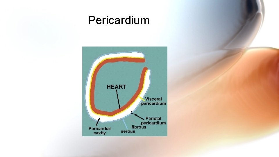
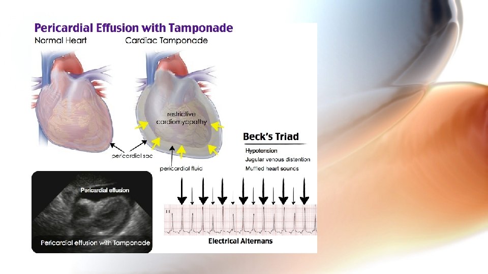
Pericardial Sac • Composed of four layers • Fibrous pericardium-tough outermost membrane • Inner membrane (serous membrane) consists of 2 layers • Parietal layer lies between the visceral layer and the fibrous pericardium • Visceral layer clings to the heart and is also known as the epicardial layer • Pericardial space • Between the visceral and the parietal layers • Contains 10 -30 ml of pericardial fluid • Minimizes friction when the heart contracts
Pericardium
Pericardial Pressure • Usually results from blood or fluid accumulation in the pericardial sac • As little as 50 -100 ml of fluid can cause a serious problem if it accumulates rapidly • A slow accumulation & increase in pressure may not produce immediate symptoms because the fibrous wall of the pericardial sac can gradually stretch to accommodate as much as 1 -2 liters of fluid
What is the effect of the pressure? • Compresses the heart • Impairs diastolic filling • Ultimately impairs diastolic filling • Compressed heart chambers • Increasing intracardiac pressure • Obstructs venous return • As blood flow into the ventricles decreases, so does cardiac output
What causes it? • Idiopathic causes such as Dressler’s syndrome • Effusion • Cancer • Bacterial infections • Tuberculosis • Hemorrhage • Trauma • Gunshot • Stab wound to chest • Penetrating chest injury
Causes, cont’d • Hemorrhage d/t nontraumatic causes • Viral or post-radiation pericarditis • Certain drug reactions • Certain connective tissue disorders
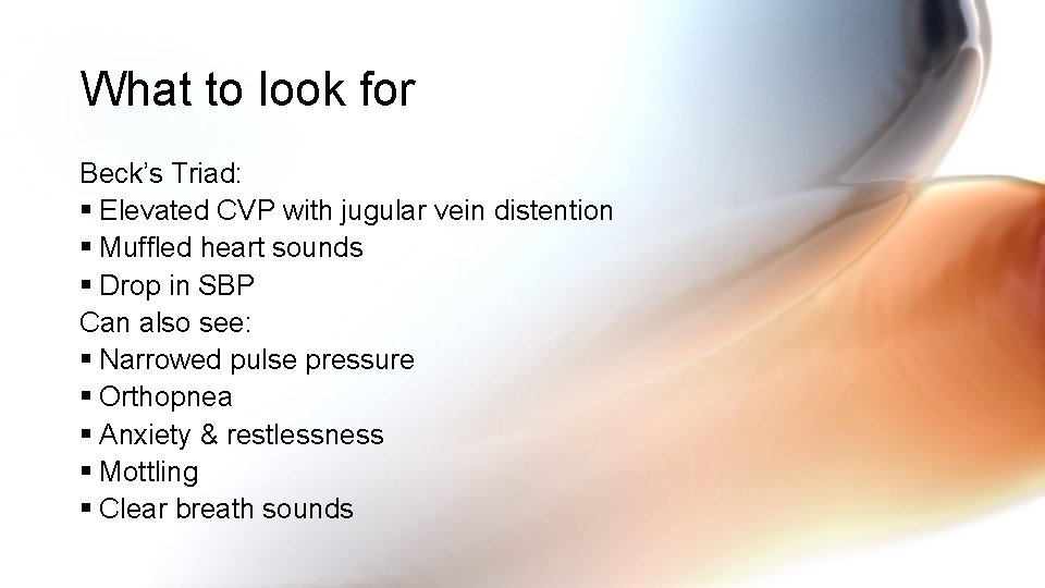
What to look for Beck’s Triad: § Elevated CVP with jugular vein distention § Muffled heart sounds § Drop in SBP Can also see: § Narrowed pulse pressure § Orthopnea § Anxiety & restlessness § Mottling § Clear breath sounds
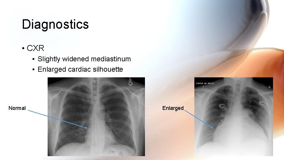
Diagnostics • CXR • Slightly widened mediastinum • Enlarged cardiac silhouette Normal Enlarged
Diagnostics, cont’d • PA catheterization reveals increased CVP; right ventricular diastolic pressure, & increased PAWP along with decreased cardiac output/cardiac index • Echo may reveal pericardial effusion with signs of right ventricular and atrial compression • CT or MRI can identify pericardial effusion or pericardial thickening
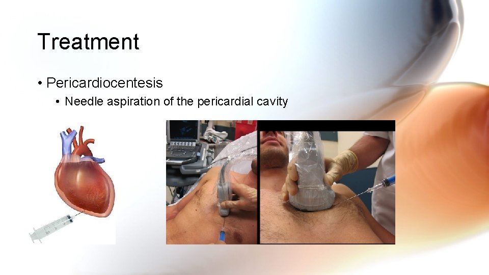
Treatment • Pericardiocentesis • Needle aspiration of the pericardial cavity
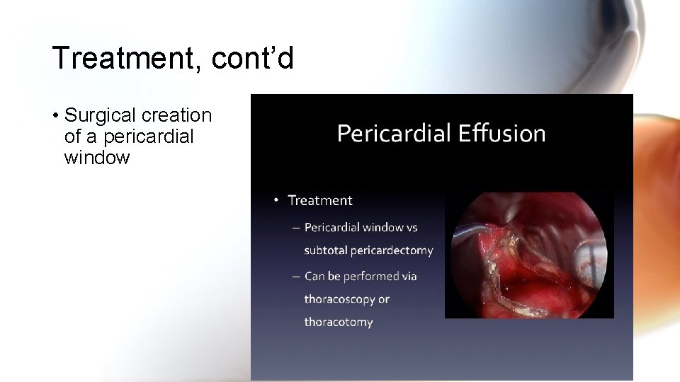
Treatment, cont’d • Surgical creation of a pericardial window
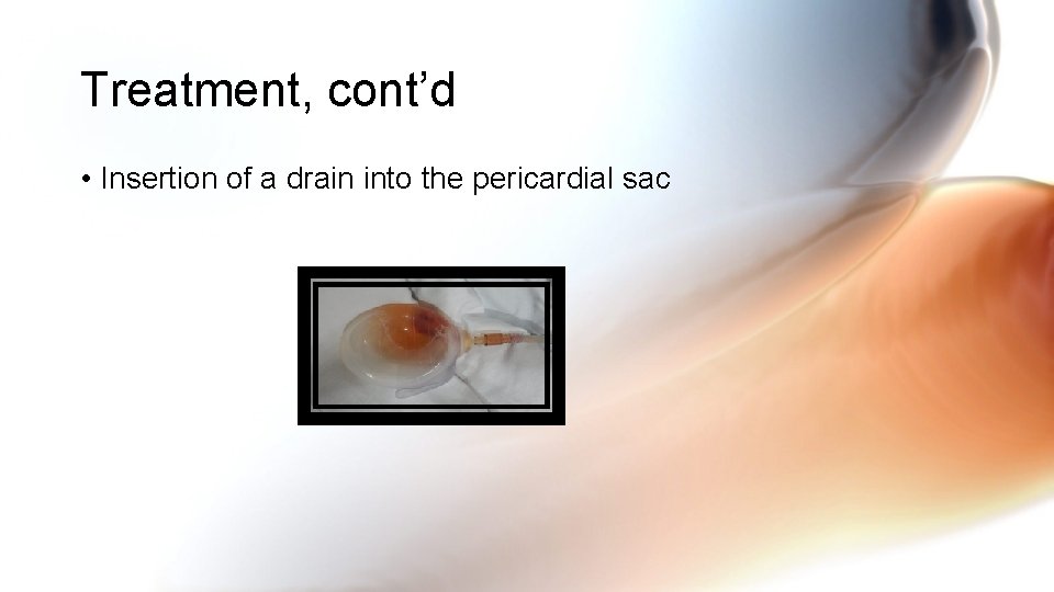
Treatment, cont’d • Insertion of a drain into the pericardial sac
Nursing Actions • Frequent CV monitoring • Assess hemodynamic status • Monitor for pulsus paradoxus • abnormally large decrease in stroke volume, systolic blood pressure and pulse wave amplitude during inspiration • Monitor for arrhythmias and ST changes • Infuse IV solutions and inotropic drugs, such as dobutamine, to maintain BP • Monitor O 2 status and patient’s airway • Prepare for pericardial procedure to remove excess pressure
Link to video https: //youtu. be/7 z. Od. P 9 u. DP 0 I I really like these Osmosis production videos