Approach to a Case of Cataract Sandeep Saxena


- Slides: 18
Approach to a Case of Cataract Sandeep Saxena MS, FRCS (Edin), FRCS (Glasg) Professor, Ophthalmology, KGMU
Differential diagnosis Painless, progressive diminution of vision • • • Cataract Primary open angle glaucoma Diabetic retinopathy Corneal dystrophies and degenerations Age related macular degeneration Retinitis pigmentosa
Cataract • Opacification of the human crystalline lens • Major cause of blindness worldwide • Classification-Etiological -Morphological
Morphological classification • Capsular cataract -Anterior -Posterior • Subcapsular cataract -Anterior -Posterior • Cortical cataract • Nuclear cataract • Polar cataract
Etiological classification I. Congenital and Developmental cataract II. Acquired cataract • Senile cataract • Traumatic cataract (blunt, penetrating, radiation, electric shock, glass blowers, infra-red) • Complicated cataract (uveitis-induced) • Metabolic cataract (Diabetes - snowflake, Wilson’s disease-sunflower) • Drug induced cataract- corticosteroids, miotics • Cataract associated with syndromes
• Congenital or Developmental cataract - Occur due to maternal infection or malnutrition, perinatal hypoxia – APH, or may be hereditary - Various morphological forms: – – – – Blue dot Sutural Fusiform or spindle shaped Embryonal nuclear Zonular Coronary Anterior or posterior polar
Senile cataract • ‘Age-related cataract’ • By the age of 70 years, over 90% of the individuals develop senile cataract • Usually bilateral, but almost always asymmetrical
Symptoms • Gradual, painless progressive loss of vision • Discomfort / glare in daylight – nuclear cataract; better vision in daylight – cortical cataract • Uniocular polyopia • Coloured halos • Black spots in front of eyes • ‘Second sight’
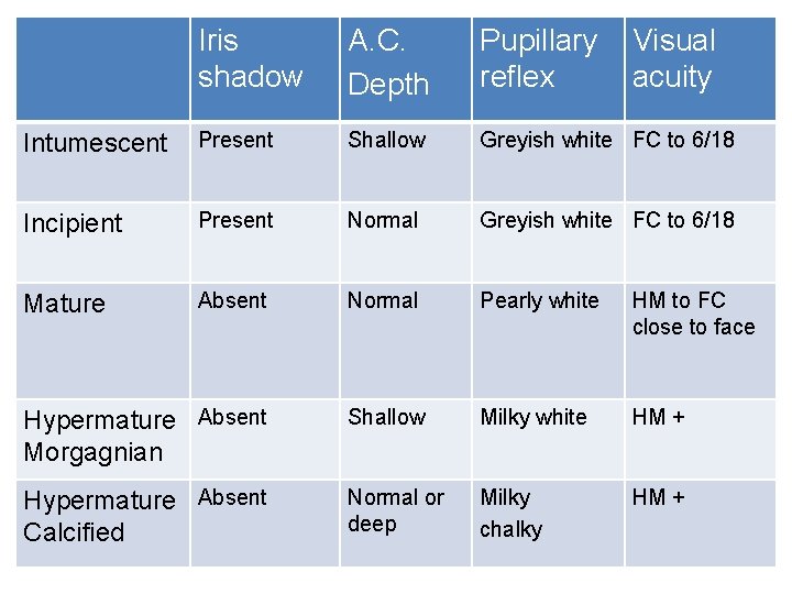
Signs • • • Iris shadow Depth of anterior chamber Pupillary reflex Visual acuity Plain mirror examination under mydriasis
Iris shadow A. C. Depth Pupillary reflex Visual acuity Intumescent Present Shallow Greyish white FC to 6/18 Incipient Present Normal Greyish white FC to 6/18 Mature Absent Normal Pearly white HM to FC close to face Hypermature Absent Morgagnian Shallow Milky white HM + Hypermature Absent Calcified Normal or deep Milky chalky HM +
Patient workup • • Retinoscopy and best corrected visual acuity Intraocular pressure Slit lamp examination Fundus evaluation – direct & indirect Macular function tests Ultrasonography IOL power calculation
General investigations • • Blood pressure Blood sugar Complete haemogram HIV, Hepatitis B & C Causes of straining Foci of infection Systemic examination
Management • An un-operated eye is more comfortable than an operated eye if visual diminution is mild. • Early cataract : -Refraction and glasses -Dark glasses or photochromatic glasses for nuclear cataract -Rule out other causes of visual diminution -If BCVA not to patient’s satisfaction, then operate.
Surgical techniques • Intracapsular cataract extraction (ICCE) • Extracapsular cataract extraction (ECCE) – Conventional ECCE – Small Incision Cataract Surgery – Phacoemulsification – Lens aspiration in paediatric (soft) cataract
Complications of cataract surgery • Intraoperative – Incision related complications – Posterior capsular rupture – Zonular dehisence – Vitreous loss – Nuclear drop – Posterior loss of lens fragments – Injury to the cornea, iris and lens – Expulsive choroidal haemorrhage
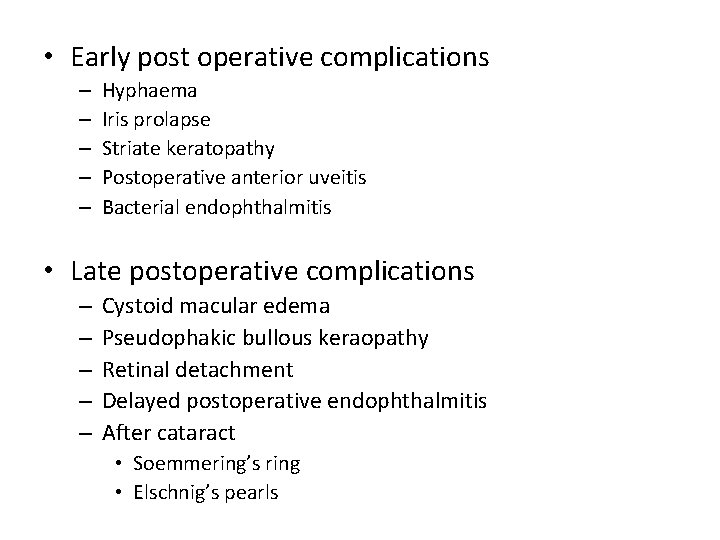
• Early post operative complications – – – Hyphaema Iris prolapse Striate keratopathy Postoperative anterior uveitis Bacterial endophthalmitis • Late postoperative complications – – – Cystoid macular edema Pseudophakic bullous keraopathy Retinal detachment Delayed postoperative endophthalmitis After cataract • Soemmering’s ring • Elschnig’s pearls
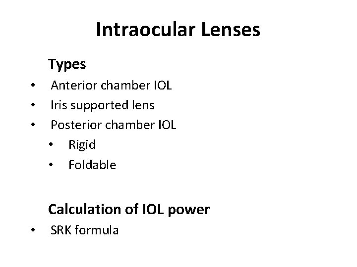
Intraocular Lenses Types • • • Anterior chamber IOL Iris supported lens Posterior chamber IOL • Rigid • Foldable Calculation of IOL power • SRK formula
Thank you