Cerebrospinal fluid CSF DR SHUBHANGI SAXENA SHMCNYS INTRODUCTION
 DR. SHUBHANGI SAXENA SHMCNYS")
Cerebrospinal fluid (CSF) DR. SHUBHANGI SAXENA SHMCNYS

INTRODUCTION • CSF is a clear, colourless, and transparent, alkaline fluid that circulates through ventricles of brain, subarachnoid space and central canal of spinal cord. • The entire cerebral cavity enclosing the brain and spinal cord has capacity of about 1600 -1700 millilitres, about 150 ml of its capacity is occupied by CSF and remainder by the brain and spinal cord.

CSF composition and properties. PROPERTIES v Volume : 130 -150 ml of which 30 -40 ml is in the ventricular system and remaining in subarachnoid space. v Rate of formation : 0. 2 - 0. 3 ml/min v Daily secretion : 500 -550 ml v Pressure (average) : in lateral lying position-130 mm H 2 O. In sitting position 200 mm H 2 O. v Specific gravity : 1. 005 v Reaction : alkaline
 148 136")
COMPOSITIONS Composition of CSF and blood CONSTITUENT CFS BLOOD Na+ (m. Eq/L) 148 136 -145 K+ (m. Eq/L) 2. 9 3. 5 -5 Cl− (m. Eq/L) 120 -130 100 -106 Ca+2 (m. Eq/L) 2. 3 4. 7 Protein (mg %) 15 -45 Glucose (mg %) 50 -75 70 -100 p. H 7. 3 7. 4

• The CSF differ from blood in having a lower concentration of K+ , glucose and protein and a higher concentration of Na+ and Cl− • CSF normally lacks blood cells.

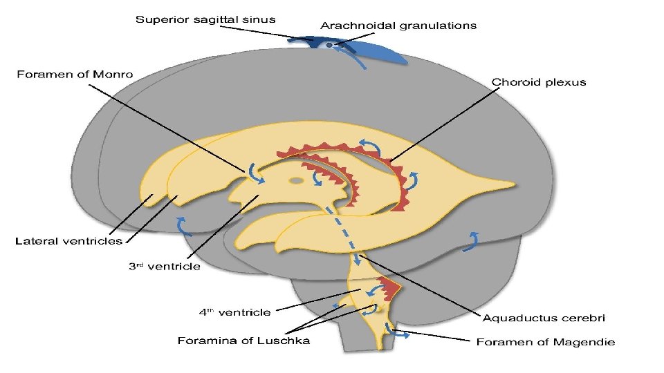
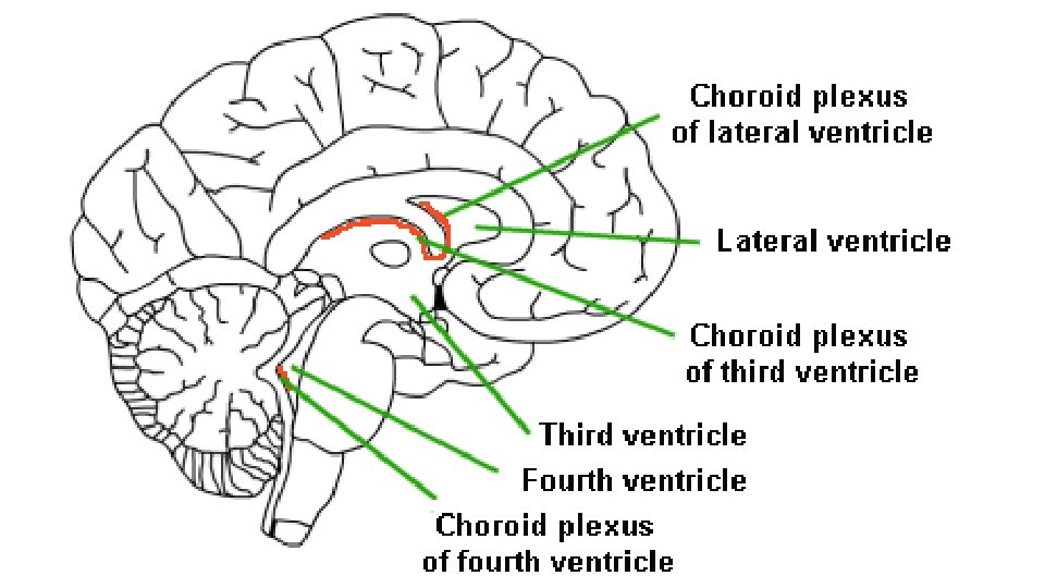
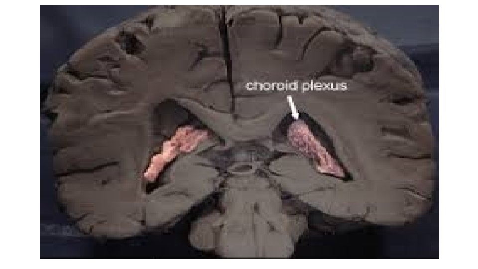
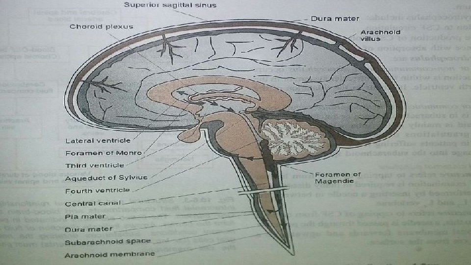
FORMATION OF CSF • 50% by choroid plexuses in the ventricles. • 40% by blood vessels of meningeal and ependymal lining of ventricles. • 10% by blood vessels of brain and spinal cord. • Mainly formed by the choroid plexus, which are covered by specialized ependymal cells. The choroid plexus are located in the cerebral ventricles ( lateral, third and fourth).

CIRCULATION OF CSF
 Into third")
CSF formed in the lateral ventricle through interventricular foramina (of monra) Into third ventricle fluid flow through cerebral aqueduct (of sylvius) Into fourth ventricle foramen of magendie, foramin of luschka Then some CSF passes into the central canal of spinal cord But most escape into the subarachnoid space (surrounding the brain and spinal cord)
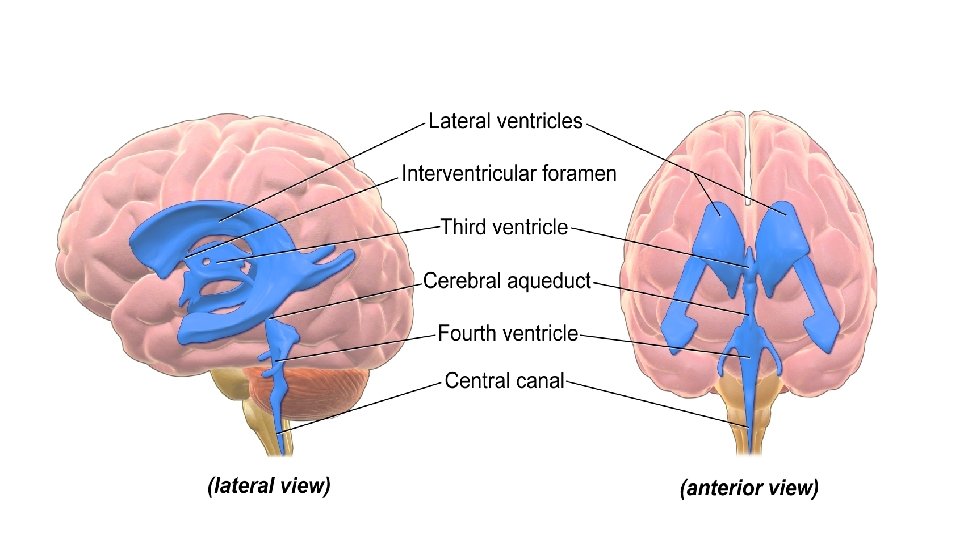

Ventricles of brain

ABSORPTION OF CSF

• Mainly 80 % by arachnoid villi into venous sinuses and spinal veins. From subarachnoid space It flows upward towards cerebrum, where almost all arachnoid villi are located and is absorbed through these villi into the cerebral venous sinuses.

FUNCTIONS OF CSF

v Protective function : CSF protects the brain from shock. The specific gravity of brain and CSF is more or less same, brain floats in CSF. When head receives a blow CSF acts like a cushion And prevent the movement of brain against the skull bone Prevent the damage of brain.

v Medium of exchange : CSF is the medium through which many substances particularly the nutritive substances and waste materials are exchanged between blood and brain tissues. v It acts as a reservoir to regulate the contents of cranium i. e it can compensate for fluctuations in the amount of blood within skull, therefore at any moment if blood volume of brain increases then CSF drains away. Conversely, if brain blood volume shrinks more CSF is retained. Thus, CSF keeps the total volume of cranial content constant. v Transport hormones and hormones releasing factors.

CLINICAL ASPECTS
 Causes of increase in CSF pressure Physiological pathological üPHYSIOLOGICAL : § Increase")
(A) Causes of increase in CSF pressure Physiological pathological üPHYSIOLOGICAL : § Increase in venous pressure for example following coughing or crying or compression of internal jugular vein. § Queckenstedt’s sign : compression of internal jugular vein decrease absorption of CSF, as a result CSF pressure increases.

ü PATHOLOGICAL : § Increase in rate of fluid formation eg: inflammation of meninges. § Increased resistance to absorption through arachnoids villi for example : brain tumors, hemorrhage or infection (cellular elements block the absorption) (B) CAUSES OF DECREASE IN CSF PRESSURE • Decrease in venous pressure • Decrease in rate of fluid formation
 The effect of I. V injection of hypertonic solutions on the CSF pressure.")
(C) The effect of I. V injection of hypertonic solutions on the CSF pressure. • I. V injection of 50 ml of 10% Na. Cl solution causes fall in CSF pressure for 2 hours due to absorption of fluid from CSF into the plasma. However effect is temporary, because Na+ and Cl− eventually move into the CSF themselves and equilibrium is re-established. • Uses : it is of value in conditions of raised intracranial pressure caused by cerebral tumors therefore helpful in : • Relieving papilloedema (optic disc swelling that is caused by increased intracranial pressure). • Restoring consciousness. • Intracranial operations made easier as bulging of the brain is prevented.
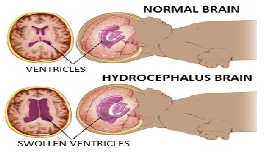
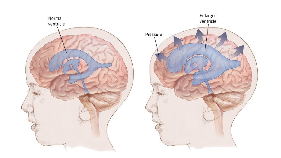
 HYDROCEPHALUS § It refers to an abnormal accumulation of CSF in the cranium.")
(D) HYDROCEPHALUS § It refers to an abnormal accumulation of CSF in the cranium. CAUSES : 1) Obstruction to CSF circulation. 2) Excessive production of CSF. 3) Interference with absorption of CSF. TYPES : Internal or non External or communicating hydrocephalus hydrocephalus

v Internal or non communicating hydrocephalus: When obstruction is within the ventricular system or in the roof of 4 th ventricle. It results in the dilatation of the ventricle. Common side of blocks are : foramina of monra, aqueduct of sylvius, foramen of magendie, foramin of luschka and within ventricular system itself. v. External or communicating hydrocephalus: Excess of fluid accumulation in subarachnoid space. Cause – when rate of CSF formation is more than rate of its absorption.

HYDROCEPHALUS

CSF Collection

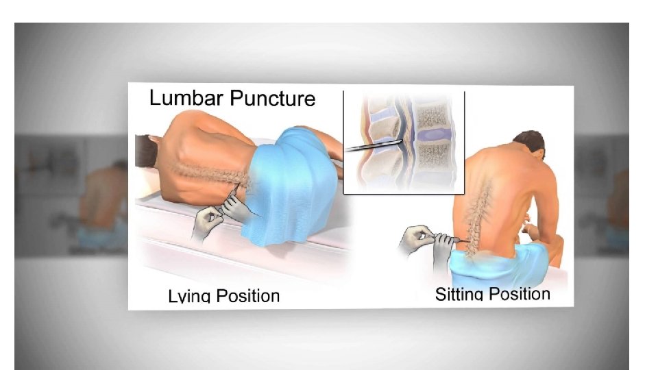
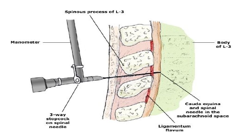
CSF Collection cisternal puncture lumbar puncture CSF is collected by passing needle is Introduce a needle between occipital into the subarachnoid bone and atlas, so that it space in the lumbar enters the cisterna magna region between the 3 rd and 4 th lumbar spine

CISTERNAL PUNCTURE

LUMBAR PUNCTURE Posture of the body : The reclining body is bent forward So as flex the vertebral column as far as possible The body is brought near the edge of the table

POSTURE

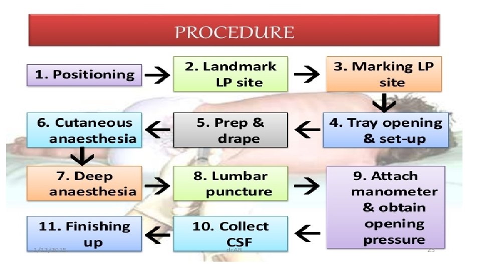
PROCEDURE The highest point of the iliac crest is determined by palpation A line is drawn on the back of the subject by joining the highest point of iliac crest of both sides Opposite the mid point, this line can cross the 4 th lumbar spine After determining the area of 4 th lumbar spine, 3 rd lumbar spine is palpated

Then the needle is introduced through the soft tissue space between the 2 spines So that the needle reaches the subarachnoid space
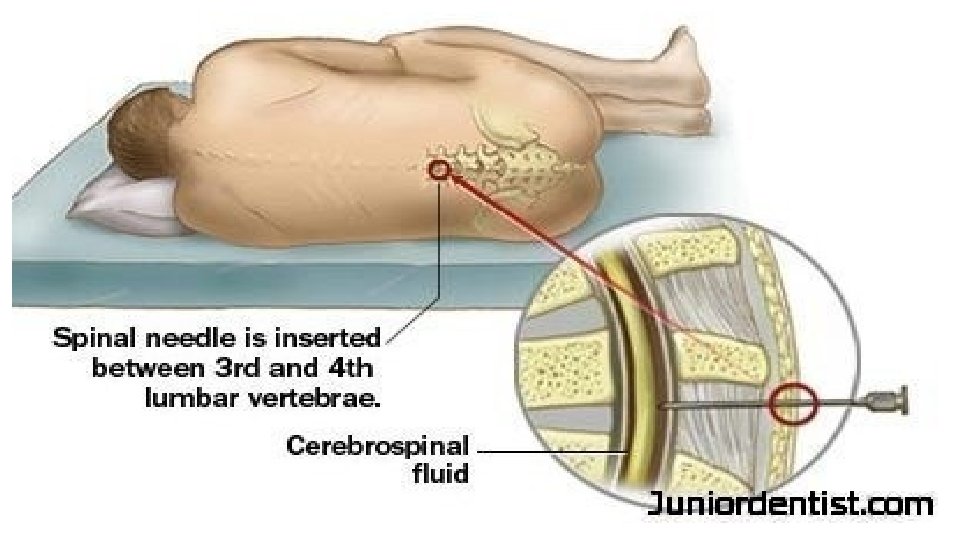

Reason for selecting the site : Ø spinal cord will not be injured because it terminates below the lower border of the 1 st lumbar vertebra The cauda equina may be damaged. Even if it is injured, it can regenerate Ø The subarachnoid space is wider in this site, it is because the pia matter is reduced very much.

Collection of CSF

USES Collecting CSF for diagnostic purpose Injecting drugs for spinal anesthesia, analgesia, chemotherapy Measuring pressure exerted by CSF

ABNORMAL CSF Ø In cerebral/ subarachnoid hemorrhage-CSF contains blood. Ø In purulent meningitis – CSF is cloudy, cell count high and consist mostly of neutrophils. chemically, the sugar concentration falls sharply and protein concentration rises. All these features are due to the acute inflammation of the meninges. ØIn tubercular meningitis, protein concentration rises but the sugar and chloride concentration falls. Ø In viral encephalitis, rise of the protein, concentration may be the only abnormality.
- Slides: 43