AlMustansiriyah University College of Medicine Department of Medicine

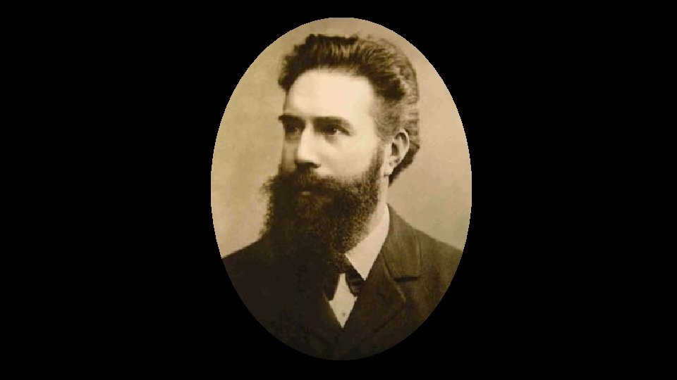
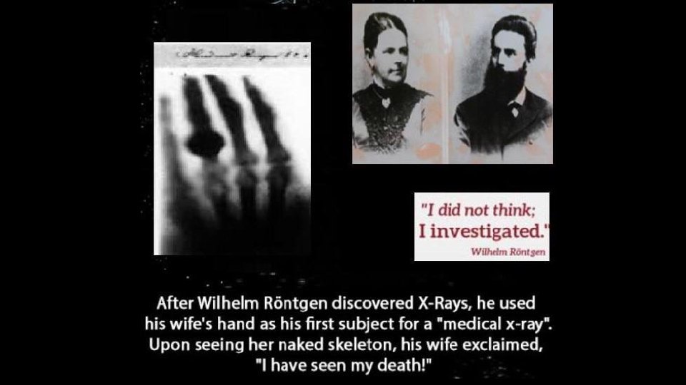
 Wilhelm Roentgen 1895 / inventor. X-rays are produced in")

 CR employs cassettes that are inserted into")


 CT scanners became available in the early 1990 s. Multidetector")


 is sensitive to the random Brownian motion")


 Scintigraphy refers to the use of gamma radiation to form images")
: The main applications of SPECT–CT include: o 99 m.")
 and PET-CT o PET is an established imaging technique, most")
: Many medical imaging departments now employ large computer")


- Slides: 34
Al-Mustansiriyah University College of Medicine - Department of Medicine Division of Radiology INTRODUCTION TO MEDICAL IMAGING DR. ABDULLATEEF AL-BAYATI Teaching Board Member CABMS-RAD 1
Conventional radiography (X-rays, plain films) Wilhelm Roentgen 1895 / inventor. X-rays are produced in an X-ray tube by focusing a beam of high-energy electrons onto a tungsten target. X-rays are a form of electromagnetic radiation, able to pass through the human body and produce an image of internal structures. The resulting image is called a radiograph. As a beam of X-rays passes through the human body, some of the X-rays are absorbed or scattered producing reduction or attenuation of the beam. Radio-opaque or radiolucent.
Five principal densities are recognized on plain radiographs, listed in order of increasing density: 1. Air/gas: black, e. g. lungs, bowel and stomach 2. Fat: dark grey, e. g. subcutaneous tissue layer, retroperitoneal fat 3. Soft tissues/water: light grey, e. g. solid organs, heart, blood vessels, muscle and fluid-filled organs such as bladder. 4. Bone: off-white 5. Contrast material/metal: bright white.
Conventional radiography Darkroom processors Computed radiography (CR) CR employs cassettes that are inserted into a laser reader following X-ray exposure. An analogue-digital converter (ADC) produces a digital image. Digital radiography (DR) DR uses a detector screen containing silicon detectors that produce an electrical signal when exposed to X-rays. This signal is analyzed to produce a digital image.
Digital radiography has many advantages over conventional radiography, including the ability to perform various manipulations on the images including: 1) 2) 3) Magnification of areas of interest Alteration of density Measurements of distances and angles.
Fluoroscopy Radiographic examination of the anatomy and motion of internal structures by a constant stream of X-rays. Uses of fluoroscopy: 1) Contrast studies of the gastrointestinal tract 2) Angiography and interventional radiology. 3) Guidance of therapeutic joint injections and arthrograms 4) Screening in theatre: operative cholangiography, retrograde pyelography, reduction and fixation of fractures, joint replacements. Fluoroscopy units fall into two categories: image intensifier and flat panel detector (FPD). FDP units have several technical advantages over image intensifier systems including smaller size, less imaging artefacts and reduced radiation exposure.
Digital subtraction angiography Digital subtraction is a process whereby a computer removes unwanted information from a radiographic image. Digital subtraction is particularly useful for angiography, referred to as DSA. Mask image contrast image subtracted image
Computed Tomography CT is an imaging technique whereby cross-sectional images are obtained with the use of X-rays. The relative density of an area of interest may be measured electronically. This density measurement is given as an attenuation value, expressed in Hounsfield units (HU) (named for Godfrey Hounsfield, the inventor of CT). • • • Water: 0 Muscle: 40 Contrast-enhanced artery: 130 Cortical bone: 500 Fat: − 50 Air: − 1000
Grey scale window width and level Mediastinal soft tissue w. Lung window Bone window
Intravenous iodinated contrast material USES in CT: Differentiation of normal blood vessels from abnormal masses, e. g. hilar vessels vs. LN. To make an abnormality more apparent, e. g. liver metastases. To demonstrate the vascular nature of a mass and thus aid in characterization. CT angiography. o o Oral contrast material is also used for abdomen CT: Differentiation of normal enhancing bowel loops from abnormal masses or fluid collections. Diagnosis of perforation of the gastrointestinal tract. Diagnosis of leaking surgical anastomoses. CT enterography. o o
Multidetector CT Helical (spiral) CT scanners became available in the early 1990 s. Multidetector row CT (MDCT)/ multislice CT (MSCT), was developed in the mid to late 1990 s. In conventional scanners, the tube and detectors rotate as the patient passes through on the scanning table. In helical CT a circular gantry holding the X-ray tube on one side and detectors on the other rotates continuously as the patient passes through. The difference with MDCT is that instead of a single row of detectors multiple detector rows are used. The original MDCT scanners used two or four rows of detectors, followed by 16 and 64 detector row scanners. At the time of writing, 256 and 320 row scanners are becoming widely available. Multidetector row CT allows the acquisition of overlapping fine sections of data, which in turn allows the reconstruction of highly accurate and detailed 3 D images as well as sections in any desired plane.
The major advantages of MDCT over conventional CT scanning are: o Increased speed of examination o Rapid examination at optimal levels of intravenous contrast concentration o Continuous volumetric nature of data allows accurate high-quality 3 D and multiplanar reconstruction.
MDCT applications including: CT angiography: coronary, cerebral, carotid, pulmonary, renal, visceral, peripheral Cardiac CT, including CT coronary angiography and coronary artery calcium scoring CT colonography (virtual colonoscopy) CT cholangiography CT enterography Brain perfusion scanning Planning of fracture repair in complex areas: acetabulum, foot, ankle and wrist. Display of complex anatomy for planning of cranial and facial reconstruction surgery o o o o
Limitations and disadvantages of CT o o Ionizing radiation. Hazards of intravenous contrast material. Lack of portability of equipment. Relatively high cost.
Ultrasound US imaging uses ultra-high-frequency sound waves to produce cross-sectional images of the body. The basic component of the US probe is the piezoelectric crystal. Hyperechoic or hypoechoic or anechoic.
US scanning is applicable to: o o o Solid organs, including liver, kidneys, spleen and pancreas. Urinary tract. Obstetrics and gynecology. Small organs including thyroid and testes. Breast. Musculoskeletal system. US-guided biopsy of various body cavities and organs. Transvaginal US (TVUS): accurate assessment of gynecological problems and of early pregnancy up to about 12 weeks’ gestation Transrectal US (TRUS): guidance of prostate biopsy; staging of rectal cancer Transcranial US: neonatal brain and TCD. Endoscopic US (EUS): assessment of tumors of the upper gastrointestinal tract and pancreas Transoesophageal echocardiography (TOE): TOE removes the problem of overlying ribs and lung, which can obscure the heart and aorta when performing conventional echocardiography.
Advantages of US over other imaging modalities include: o Lack of ionizing radiation, a particular advantage in pregnancy and pediatrics. o Relatively low cost. o Portability of equipment. Doppler US o It includes color, power, spectral and continuous wave Doppler imaging. o The Doppler effect has been applied to US imaging. Flowing blood causes an alteration to the frequency of sound waves returning to the US probe. o Color Doppler is used in many areas of US including echocardiography and vascular US. Color Doppler is also used to confirm blood flow within organs (e. g. testis to exclude torsion) and to assess the vascularity of tumors. Contrast-enhanced US o The accuracy of US in certain applications may be enhanced by the use of intravenously injected microbubble contrast agents. o Contrast-enhanced US applications: Echocardiography and assessment of liver masses Disadvantages and limitations of US o US is highly operator dependent. o US cannot penetrate gas or bone.
Magnetic Resonance Imaging MRI uses the magnetic properties of spinning hydrogen atoms to produce images. Large powerful magnet: Most current medical MRI machines have field strengths of 1. 5 or 3. 0 tesla. The hydrogen atoms within the patient align in a direction either parallel or antiparallel to the strong external field. A greater proportion aligns in the parallel direction so that the net vector of their alignment, and therefore the net magnetic vector, will be in the direction of the external field. This is known as longitudinal magnetization. • A second magnetic field is applied at right angles to the original external field. This second magnetic field is known as the radiofrequency pulse (RF pulse), because it is applied at a frequency in the same part of the electromagnetic spectrum as radio waves. A magnetic coil, known as the RF coil, applies the RF pulse. The RF pulse causes the net magnetization vector of the hydrogen atoms to turn towards the transverse plane. The component of the net magnetization vector in the transverse plane induces an electrical current in the RF coil. This current is known as the MR signal.
• • High Signal = Hyperintense Low Signal = Hypointense Intermediate Signal = Isointense Signal Void= No Signal
Tissue contrast and imaging sequences: Much of the complexity of MRI arises from the fact that the MR signal depends on many varied properties of the tissues and structures being examined, including: o o o Number of hydrogen atoms present in tissue (proton density) Chemical environment of the hydrogen atoms, e. g. whether in free water or bound by fat Flow: blood vessels or CSF Magnetic susceptibility T 1 relaxation time T 2 relaxation time. By altering the duration and amplitude of the RF pulse, as well as the timing and repetition of its application, various imaging sequences use these properties to produce image contrast. Terms used to describe the different types of MR imaging sequences include spin echo, inversion recovery and gradient-recalled echo (gradient echo).
Spin echo sequences o Spin echo sequences include T 1 -weighted, T 2 - weighted and proton density. o Following the application of a 90° RF pulse, the net magnetization vector lies in the transverse plane. Also, all of the hydrogen protons are ‘in phase’, i. e. spinning at the same rate. Upon cessation of the RF pulse, two things begin to happen: Net magnetization vector rotates back to the longitudinal direction: longitudinal or T 1 relaxation Hydrogen atoms dephase (spin at slightly varying rates): transverse or T 2 relaxation (decay).
Inversion recovery sequences are used to suppress unwanted signals that may obscure pathology. The two most common inversion recovery sequences are used to suppress fat (STIR) and water (FLAIR). Gradient- echo sequences are extremely sensitive to the presence of substances that cause local alterations in magnetic properties. Examples of such substances include iron-containing haemosiderin and ferritin found in chronic blood. It has known as susceptibility weighted imaging.
Functional MRI sequences o Diffusion-weighted imaging (DWI) is sensitive to the random Brownian motion (diffusion) of water molecules within tissue. The greater the amount of diffusion, the greater the signal loss on DWI. Areas of reduced water molecule diffusion show on DWI as relatively high signal. Diffusion-weighted imaging is the most sensitive imaging test available for the diagnosis of acute cerebral infarction, epidermoid cyst and abscess. o Perfusion-weighted imaging (PWI) may be used in patients with cerebral infarct to map out areas of brain at risk of ischemia that may be salvageable with thrombolysis. o Magnetic resonance spectroscopy (MRS) uses different frequencies to identify certain molecules in a selected volume of tissue, known as a voxel. Following data analysis, a spectrographic graph of certain metabolites is drawn. Metabolites of interest include lipid, lactate, NAA (N-acetylaspartate), choline, creatinine, citrate and myoinositol. Uses of MRS include characterization of metabolic brain disorders in children, imaging of dementias, differentiation of recurrent cerebral tumor from radiation necrosis, and diagnosis of prostatic carcinoma.
o Magnetic resonance angiography and magnetic resonance venography used for visualization of blood vessels with or without using contrast material. o Contrast material in MRI Gadolinium (Gd) is a paramagnetic substance that causes T 1 shortening and therefore increased signal on T 1 -weighted images.
Applications of MRI: o Imaging modality of choice for most brain and spine disorders o Musculoskeletal disorders, including internal derangements of joints and staging of musculoskeletal tumors o Cardiac MR is an established technique in specific applications including assessment of congenital heart disease and aortic disorders o MR of the abdomen is used in adults for visualization of the biliary system, and for characterization of hepatic, renal, adrenal and pancreatic tumors o In children, MR of the abdomen is increasingly replacing CT for the diagnosis and staging of abdominal tumors o MRA is widely used in the imaging of the cerebral circulation and in some centers is the initial angiographic method of choice for other areas including the renal and peripheral circulations.
Particular advantages of MRI include: o o Excellent soft tissue contrast and characterization Lack of artefact from adjacent bones, e. g. pituitary fossa Multiplanar capabilities Lack of ionizing radiation. Disadvantages and limitations of MRI o o o o Time taken to complete examination. Young children and infants usually require general anesthesia Patients experiencing pain may require intravenous pain relief For examination of the abdomen, an antispasmodic, such as intravenous hyoscine, may be required to reduce movement of the bowel Safety issues related to ferromagnetic materials within the patient, e. g. surgical clips, or electrical devices such as pacemakers. High auditory noise levels: earplugs should be provided to all patients undergoing MRI examinations Claustrophobia Adverse contrast (gadolinium) reaction: allergy and nephrogenic systemic fibrosis (rare).
Scintigraphy (nuclear medicine) Scintigraphy refers to the use of gamma radiation to form images following the injection of various radiopharmaceuticals. The gamma rays emitted by the radionuclides are detected by a gamma camera that converts the absorbed energy of the radiation to an electric signal. This signal is analysed by a computer and displayed as an image.
Single photon emission CT (SPECT): The main applications of SPECT–CT include: o 99 m. Tc-MDP bone scanning o 201 Tl cardiac scanning o 99 m. Tc-MIBG staging of neuroblastoma o Cerebral perfusion studies.
Positron emission tomography (PET) and PET-CT o PET is an established imaging technique, most commonly used in oncology. o The most commonly used radiopharmaceutical in PET scanning is FDG (2 deoxyglucose labelled with the positron-emitter fluorine-18). o The current roles of PET imaging may be summarized as follows: Oncology: Tumor staging. Assessment of tumor response to therapy. Differentiate benign and malignant masses, e. g. solitary pulmonary nodule. Detect tumor recurrence. Cardiac: Non-invasive assessment of myocardial viability in patients with coronary artery disease. Central nervous system: Characterization of dementia disorders. Localization of seizure focus in epilepsy.
Picture archiving and communication systems (PACS): Many medical imaging departments now employ large computer storage facilities and networks known as picture archiving and communication systems (PACS). Images obtained by CR and DR are stored digitally, as are images from other modalities including computed tomography (CT), magnetic resonance imaging (MRI), ultrasound (US) and scintigraphy. PACS systems allow instant recall and display of a patient’s imaging studies. Images can be displayed on monitors throughout the hospital in wards, meeting rooms and operating theatres as required.
Q 1. Define the Hounsfield unit and describe its value. Q 2. What means by CR and DR? and what is the difference between them.