Action Potential II the HodgkinHuxley Axon Hodgkin and


- Slides: 18

Action Potential – II (the Hodgkin-Huxley Axon) • • Hodgkin and Huxley 1952 papers membrane permeability during AP AP threshold AP propagation

Fig. 2. 1

Voltage clamp Hypothesis: potential-sensitive Na+ and K+ permeability changes are both necessary and sufficient for the production of action potentials.

Voltage dependent permeabilities. Fig 3. 1

What ionic species are engaged? Fig 3. 2

The I/V curve of an AP Fig 3. 3

The involvement of Na+ (and K+) Fig 3. 4

Two separate ionic conductances (TEA) (TTX) Fig 3. 5

A rigorous description of membrane conductances V = IR (Ohm’s law) for Na+ in neurons: V = Vm - ENa (“driving force”, fixed in v-clamp) I = ionic current R = 1/conductance, g therefore, g = I/V

Na and K conductances with time Both conductances are voltage- and time-dependent g. Na+ quickly activates, then inactivates g. K+ slowly activates, does not inactivate Fig 3. 6

Na+ and K+ conductance are voltage dependent Na+ channel activation defines threshold fig 3. 7

Mathematical reconstruction of the AP g. Na+ and g. K+ fully explain AP: - shape - threshold - after hyperpolarization - refractory period - propagation Fig. 3. 8

Passive conductance is not great in an axon Fig 3. 10

Passive membrane properties Ch. 3 Box C

Propagation of an action potential Fig 3. 11

The mechanism of action potential conduction Fig 3. 12

AP propagation in a myelinated axon (“saltatory”) Fig 3. 13

Ion channel distribution in nodes of Ranvier Na+ channels in node of Ranvier (Rasband Shragger 2000)
 Difference hodgkin and non hodgkin lymphoma
 Diff between hodgkin and non hodgkin
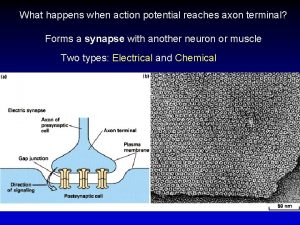
 What happens when action potential reaches axon terminal
 Graded potential and action potential
 Neuronal pool
 Define graded potential
 Graded vs action potential
 Refractory period in action potential
 Sources of bioelectric potentials
 Hypopolarization
 End plate potential
 Axon hillock
 Action potential resting potential
 Define electric potential and potential difference.
 Volts to ev
 Water potential
 Difference between sales potential and market potential
 Electric potential and potential difference
 Joules per coloumb
