Myelinated Motor Axon Soma Axon Hillock Synapse Myelin


- Slides: 11
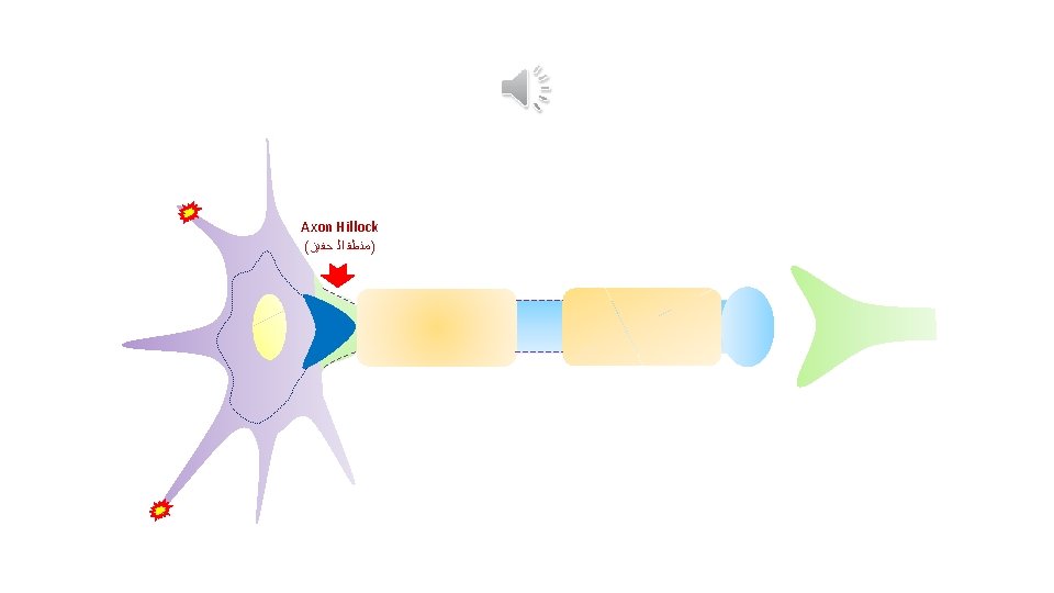
Myelinated Motor Axon Soma Axon Hillock Synapse Myelin Sheath 1 st Node of Ranvier Internodal Segment At rest, the microtubules’ montage protrudes into the lumen of the Axon Hillock The dashed line indicates The interrupted line indicates the massive presence of Na+ pressure-gated channels Na+ pressure- gated channels in cell membrane in the membrane of Ranvier node of Axon Hillock Intraluminal Resting Pressure At rest as well as in action, Neurotransmitters are always present in Synaptic Cleft
The microtubules' montage contracts and then retracts backward into the Soma Resting Pressure A zone of negative pressure (Trough) is built up within the lumen of Axon Hillock
A central pressure wave is built up and spreads toward the cell membrane of Soma Cell Membrane Resting Pressure Microtubules’ Network A zone of negative pressure (Trough) is built up within the lumen of Axon Hillock
The microtubules’ montage relaxes and then is briskly pushed forward to its initial position The central pressure wave rebounds off cell membrane and comes back to the point of starting A zone of Positive pressure (Crest) is built up within the lumen of Axon Hillock Resting Pressure
1 st. Ca Node ++ of Ranvier Preliminary Action Pressure Wave Crest Trough
++ Ca Standard Action Pressure Wave Postsynaptic Dendrite Resting Pressure Crest Trough
1 st. Ca Node of Ranvier ++