Update on VARC Patrick W Serruys Nicolo Piazza

- Slides: 23
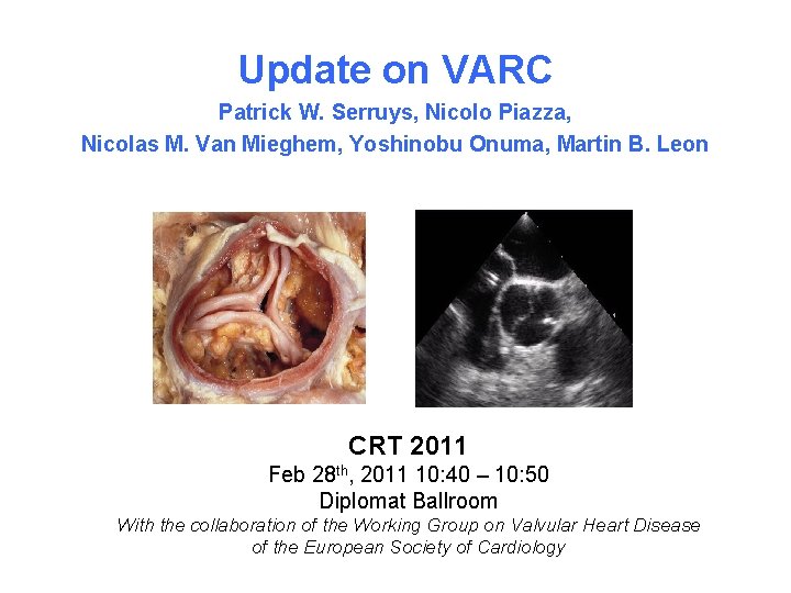
Update on VARC Patrick W. Serruys, Nicolo Piazza, Nicolas M. Van Mieghem, Yoshinobu Onuma, Martin B. Leon CRT 2011 Feb 28 th, 2011 10: 40 – 10: 50 Diplomat Ballroom With the collaboration of the Working Group on Valvular Heart Disease of the European Society of Cardiology
Patrick W. Serruys, MD, Ph. D I have no real or apparent conflicts of interest to report.
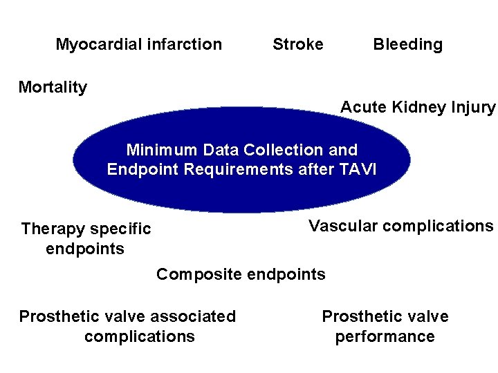
Myocardial infarction Stroke Bleeding Mortality Acute Kidney Injury Minimum Data Collection and Endpoint Requirements after TAVI Vascular complications Therapy specific endpoints Composite endpoints Prosthetic valve associated complications Prosthetic valve performance
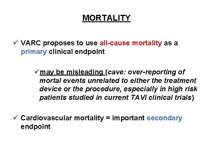
MORTALITY ü VARC proposes to use all-cause mortality as a primary clinical endpoint ümay be misleading (cave: over-reporting of mortal events unrelated to either the treatment device or the procedure, especially in high risk patients studied in current TAVI clinical trials) ü Cardiovascular mortality = important secondary endpoint
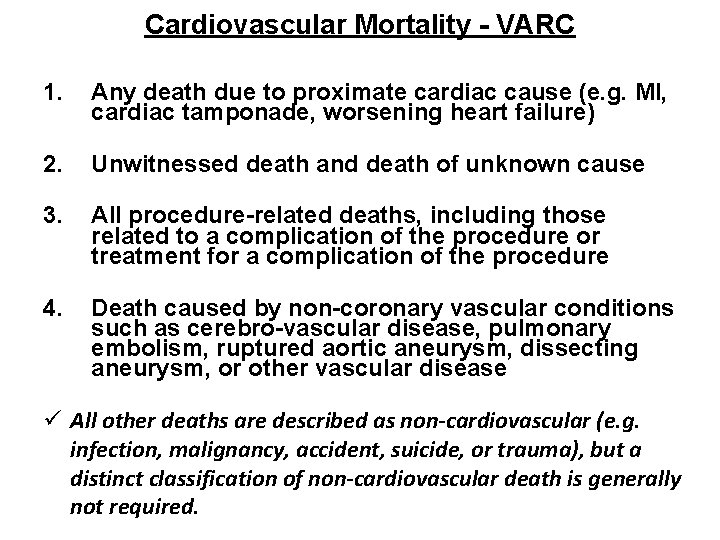
Cardiovascular Mortality - VARC 1. Any death due to proximate cardiac cause (e. g. MI, cardiac tamponade, worsening heart failure) 2. Unwitnessed death and death of unknown cause 3. All procedure-related deaths, including those related to a complication of the procedure or treatment for a complication of the procedure 4. Death caused by non-coronary vascular conditions such as cerebro-vascular disease, pulmonary embolism, ruptured aortic aneurysm, dissecting aneurysm, or other vascular disease ü All other deaths are described as non-cardiovascular (e. g. infection, malignancy, accident, suicide, or trauma), but a distinct classification of non-cardiovascular death is generally not required.
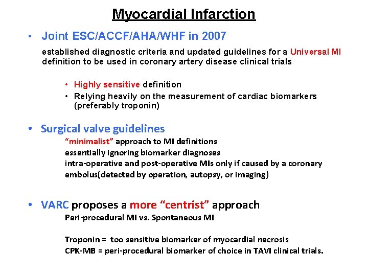
Myocardial Infarction • Joint ESC/ACCF/AHA/WHF in 2007 established diagnostic criteria and updated guidelines for a Universal MI definition to be used in coronary artery disease clinical trials • Highly sensitive definition • Relying heavily on the measurement of cardiac biomarkers (preferably troponin) • Surgical valve guidelines “minimalist” approach to MI definitions essentially ignoring biomarker diagnoses intra-operative and post-operative MIs only if caused by a coronary embolus(detected by operation, autopsy, or imaging) • VARC proposes a more “centrist” approach Peri-procedural MI vs. Spontaneous MI Troponin = too sensitive biomarker of myocardial necrosis CPK-MB = peri-procedural biomarker of choice in TAVI clinical trials.
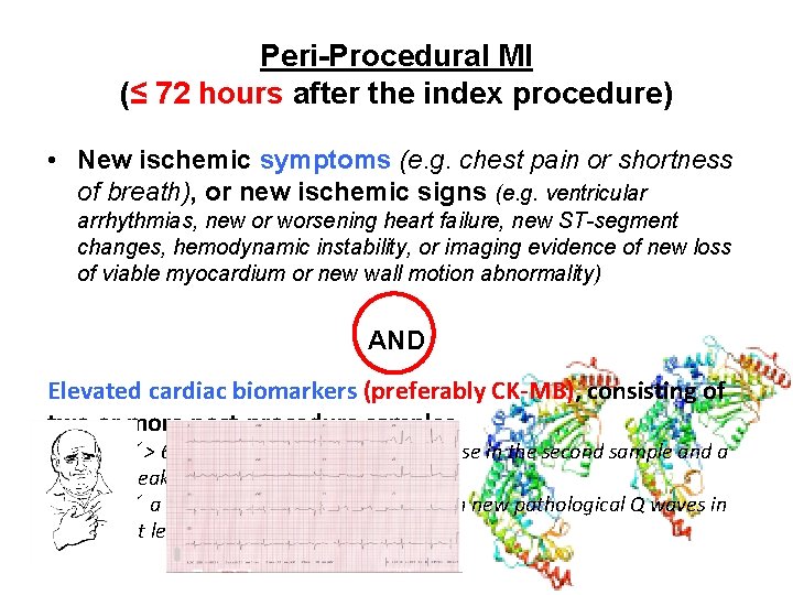
Peri-Procedural MI (≤ 72 hours after the index procedure) • New ischemic symptoms (e. g. chest pain or shortness of breath), or new ischemic signs (e. g. ventricular arrhythmias, new or worsening heart failure, new ST-segment changes, hemodynamic instability, or imaging evidence of new loss of viable myocardium or new wall motion abnormality) AND Elevated cardiac biomarkers (preferably CK-MB), consisting of two or more post-procedure samples ü> 6 -8 hours apart with a 20% increase in the second sample and a peak value exceeding 10 x URL, or ü a peak value exceeding 5 x URL with new pathological Q waves in at least 2 contiguous leads.
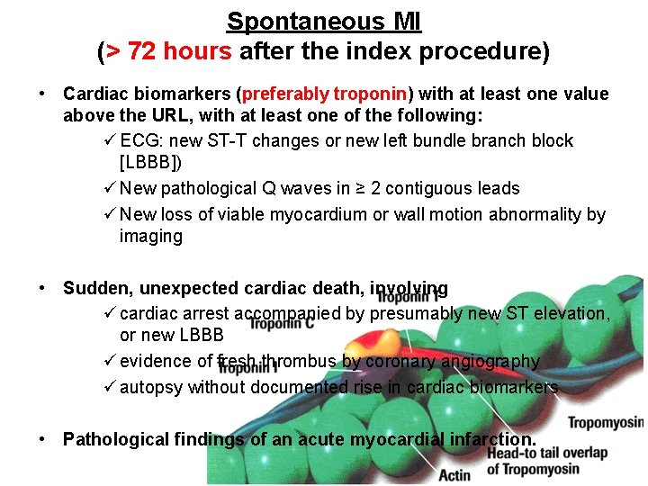
Spontaneous MI (> 72 hours after the index procedure) • Cardiac biomarkers (preferably troponin) with at least one value above the URL, with at least one of the following: ü ECG: new ST-T changes or new left bundle branch block [LBBB]) ü New pathological Q waves in ≥ 2 contiguous leads ü New loss of viable myocardium or wall motion abnormality by imaging • Sudden, unexpected cardiac death, involving ü cardiac arrest accompanied by presumably new ST elevation, or new LBBB ü evidence of fresh thrombus by coronary angiography ü autopsy without documented rise in cardiac biomarkers • Pathological findings of an acute myocardial infarction.
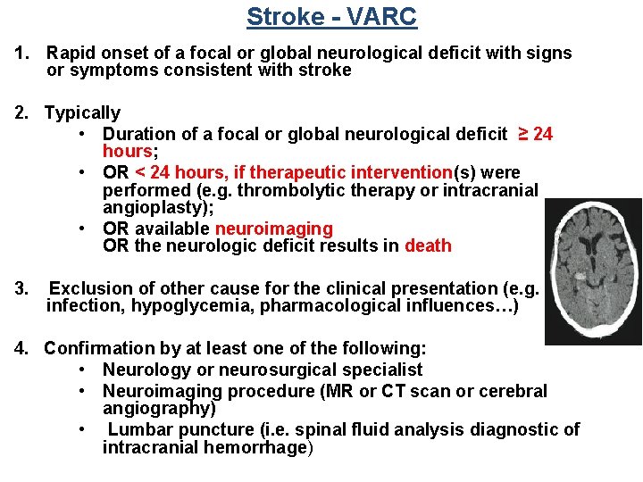
Stroke - VARC 1. Rapid onset of a focal or global neurological deficit with signs or symptoms consistent with stroke 2. Typically • Duration of a focal or global neurological deficit ≥ 24 hours; • OR < 24 hours, if therapeutic intervention(s) were performed (e. g. thrombolytic therapy or intracranial angioplasty); • OR available neuroimaging OR the neurologic deficit results in death 3. Exclusion of other cause for the clinical presentation (e. g. infection, hypoglycemia, pharmacological influences…) 4. Confirmation by at least one of the following: • Neurology or neurosurgical specialist • Neuroimaging procedure (MR or CT scan or cerebral angiography) • Lumbar puncture (i. e. spinal fluid analysis diagnostic of intracranial hemorrhage)
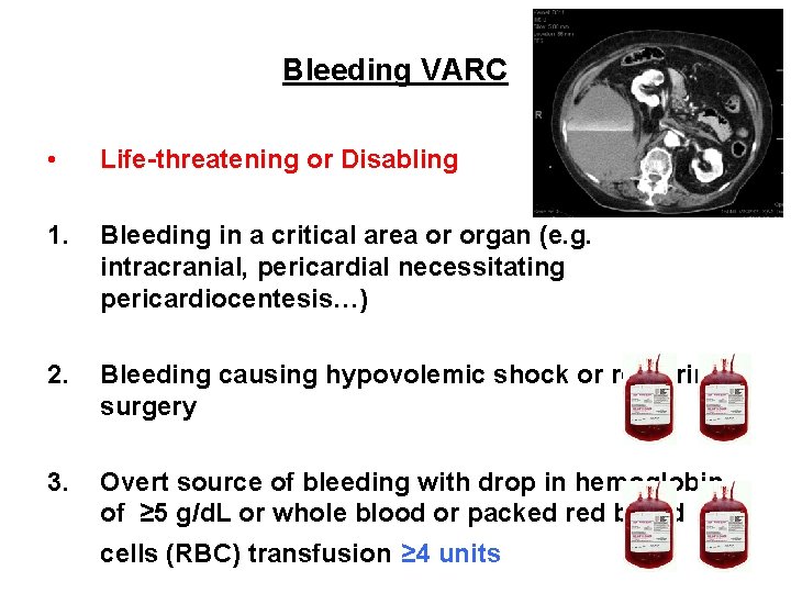
Bleeding VARC • Life-threatening or Disabling 1. Bleeding in a critical area or organ (e. g. intracranial, pericardial necessitating pericardiocentesis…) 2. Bleeding causing hypovolemic shock or requiring surgery 3. Overt source of bleeding with drop in hemoglobin of ≥ 5 g/d. L or whole blood or packed red blood ≥ 4 cells (RBC) transfusion ≥ 4 units
Bleeding VARC 2 -3 Unit • Major 1. Overt bleeding either 1. with a drop in the hemoglobin level of at least 3. 0 g/d. L 2. requiring transfusion of 2 -3 units of whole blood/RBC • Minor Any bleeding worthy of clinical mention (e. g. access site hematoma) that does not qualify as lifethreatening, disabling or major
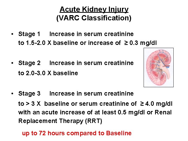
Acute Kidney Injury (VARC Classification) • Stage 1 Increase in serum creatinine to 1. 5 -2. 0 X baseline or increase of ≥ 0. 3 mg/dl • Stage 2 Increase in serum creatinine to 2. 0 -3. 0 X baseline • Stage 3 Increase in serum creatinine to > 3 X baseline or serum creatinine of ≥ 4. 0 mg/dl with an acute increase of at least 0. 5 mg/dl or Renal Replacement Therapy (RRT) up to 72 hours compared to Baseline
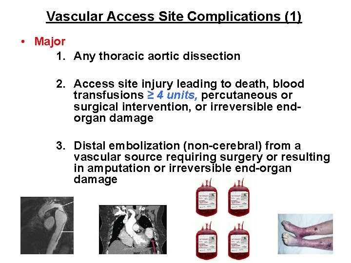
Vascular Access Site Complications (1) • Major 1. Any thoracic aortic dissection 2. Access site injury leading to death, blood transfusions ≥ 4 units, percutaneous or surgical intervention, or irreversible endorgan damage 3. Distal embolization (non-cerebral) from a vascular source requiring surgery or resulting in amputation or irreversible end-organ damage
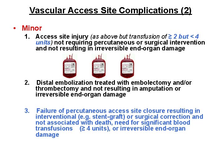
Vascular Access Site Complications (2) • Minor 1. Access site injury (as above but transfusion of ≥ 2 but < 4 units) not requiring percutaneous or surgical intervention and not resulting in irreversible end-organ damage 2. Distal embolization treated with embolectomy and/or thrombectomy and not resulting in amputation or irreversible end-organ damage ≥ 4 3. Failure of percutaneous access site closure resulting in interventional (e. g. stent-graft) or surgical correction and not associated with death, need for significant blood transfusions (≥ 4 units), or irreversible end-organ damage
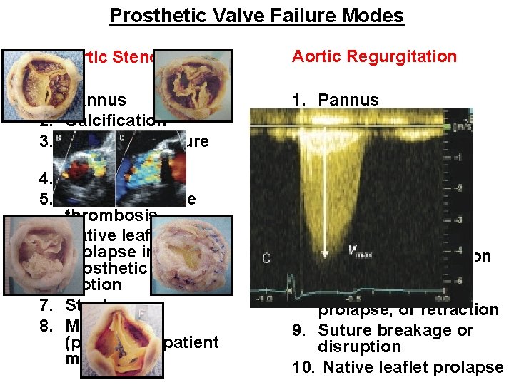
Prosthetic Valve Failure Modes Aortic Stenosis 1. Pannus 2. Calcification 3. Support structure deformation 4. Endocarditis 5. Prosthetic valve thrombosis 6. Native leaflet prolapse impeding prosthetic leaflet motion 7. Stent creep 8. Mal-sizing (prosthesis-patient mismatch) Aortic Regurgitation 1. Pannus 2. Calcification 3. Support structure deformation 4. Endocarditis 5. Prosthetic valve thrombosis 6. Malposition 7. Acute mal-coaptation 8. Leaflet wear, tear/perforation, prolapse, or retraction 9. Suture breakage or disruption 10. Native leaflet prolapse
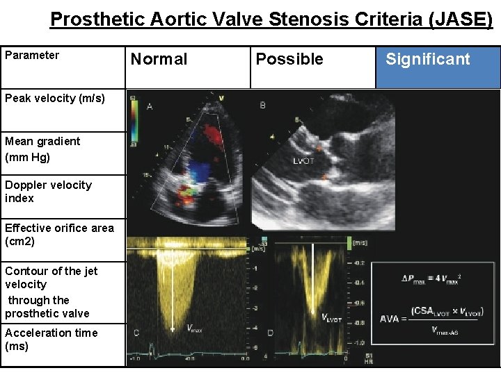
Prosthetic Aortic Valve Stenosis Criteria (JASE) Parameter Normal Possible Significant Peak velocity (m/s) <3 3 -4 >4 Mean gradient (mm Hg) <20 20 -35 >35 Doppler velocity index ≥ 0. 30 0. 29 -0. 25 <0. 25 Effective orifice area (cm 2) >1. 2 0. 8 -1. 2 <0. 8 Contour of the jet velocity through the prosthetic valve Triangular, early peaking Triangular to intermediate Rounded, symmetrical contour Acceleration time (ms) <80 80 -100 >100
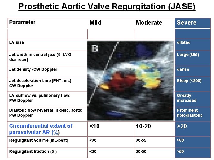
Prosthetic Aortic Valve Regurgitation (JASE) Parameter Mild Moderate Severe LV size Normal/mildly dilated Jet width in central jets (% LVO diameter) Narrow (<25%) Intermediate (2664%) Large (≥ 65) Jet density /CW Doppler Incomplete/faint Dense dense Jet deceleration time (PHT, ms) CW Doppler Slow (>500) Variable (200 -500) Steep (<200) LV outflow vs. pulmonary flow: PW Doppler Slightly increased Intermediate Greatly increased Diastolic flow reversal in desc. aorta: PW Doppler Abent or brief early diastolic Intermediate Prominent, holodiastolic Circumferential extent of paravalvular AR (%) <10 10 -20 >20 Regurgitant volume (m. L/beat) <30 30 -59 >60 Regurgitant fraction (%) <30 30 -50 >50
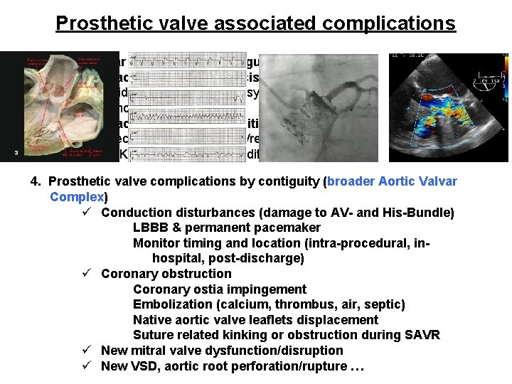
Prosthetic valve associated complications 1. Paravalvular prosthetic aortic regurgitation 2. Prosthetic aortic valve thrombosis ü Incidental finding at autopsy/reoperation ü Echocardiography 3. Prosthetic aortic valve endocarditis ü Infection focus at autopsy/reoperation ü DUKE criteria for endocarditis if no reoperation 4. Prosthetic valve complications by contiguity (broader Aortic Valvar Complex) ü Conduction disturbances (damage to AV- and His-Bundle) LBBB & permanent pacemaker Monitor timing and location (intra-procedural, inhospital, post-discharge) ü Coronary obstruction Coronary ostia impingement Embolization (calcium, thrombus, air, septic) Native aortic valve leaflets displacement Suture related kinking or obstruction during SAVR ü New mitral valve dysfunction/disruption ü New VSD, aortic root perforation/rupture …
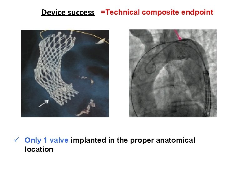
Device success =Technical composite endpoint ü Successful vascular access, delivery, device deployment and delivery system retrieval ü Correct device position in the proper anatomical location ü Intended performance of the prosthetic heart valve (aortic valve area > 1. 2 cm 2 and mean aortic valve gradient < 20 mm. Hg, without moderate or severe prosthetic valve AR) ü Only 1 valve implanted in the proper anatomical location
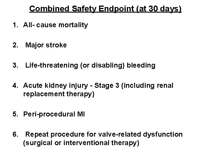
Combined Safety Endpoint (at 30 days) 1. All- cause mortality 2. Major stroke 3. Life-threatening (or disabling) bleeding 4. Acute kidney injury - Stage 3 (including renal replacement therapy) 5. Peri-procedural MI 6. Repeat procedure for valve-related dysfunction (surgical or interventional therapy)
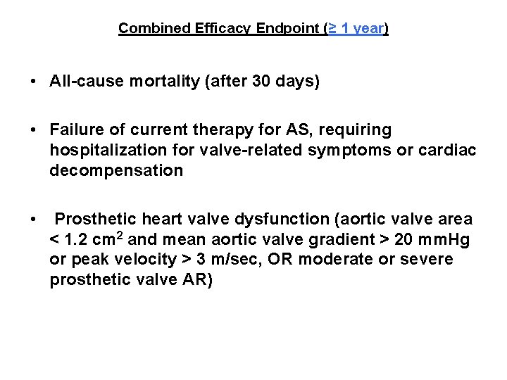
Combined Efficacy Endpoint (≥ 1 year) • All-cause mortality (after 30 days) • Failure of current therapy for AS, requiring hospitalization for valve-related symptoms or cardiac decompensation • Prosthetic heart valve dysfunction (aortic valve area < 1. 2 cm 2 and mean aortic valve gradient > 20 mm. Hg or peak velocity > 3 m/sec, OR moderate or severe prosthetic valve AR)
VARC online January 6 - 7 2011