TRAUMATOLOGY COMMONWEALTH OF AUSTRALIA Copyright Regulations 1969 WARNING





- Slides: 42
TRAUMATOLOGY
COMMONWEALTH OF AUSTRALIA Copyright Regulations 1969 WARNING This material has been reproduced and communicated to you by or on behalf of The University of Adelaide under Part VB of the Copyright Act 1968 (the Act). The material in this communication may be subject to copyright under the Act. Any further reproduction or communication of this material by you may be the subject of copyright protection under the Act. The AUDSS assumes no responsibility or liability for any information, materials or other content provided by any of our student lecturers. All content is viewed and used by you at your own risk and we do not warrant the accuracy or reliability of any of the lecture material. The views expressed are those of the individual contributors and not necessarily those of the AUDSS of Adelaide School of Dentistry. Do not remove this notice.
Tooth Discoloration Red/Brown Discoloration - Trauma → rupture of blood vessels → extravasation of erythrocytes into pulpal space Colour changes in the tooth: - Pink → yellow-pink → gray-brown-violet - Haemoglobin or breakdown products Return of tooth to normal colour = pulpal survival Pulp does not return to normal colour = one sign of pulpal necrosis Yellow Discoloration - Laying down of tertiary dentine following traumatic injury Coupled with radiographic root canal stenosis Indicates a vital pulp
Pulpal Diagnosis of Traumatised Teeth Very important - Over-diagnosis = unnecessary RCT - Failure to diagnose = untreated infection, loss of tooth structure to resorption, poorer long term prognosis Regular followup and monitoring important for traumatised teeth - Often followup of up to 5 years recommended for traumatised teeth, can have late complications - Testing at followup visits includes: pulp testing, TTP, PA radiograph, etc.
Pulpal Diagnosis of Traumatised Teeth Signs of Pulpal Healing - Transient apical breakdown: widening of the apical foramen to allow proliferation of blood vessels Slight, well-defined RL: takes several months to resolve - Root canal stenosis: deposition of tooth structure in canal - Return of normal pulp sensibility: takes minimum of 4 -6 weeks following traumatic event Signs of Pulpal Pathology - Classic signs of pulpal necrosis + apical periodontitis: negative pulp tests, TTP, discoloration, apical R/L - Internal or external root resorption - Diagnosis of pulpal pathology made = RCT Some conditions have poor prognosis (eg. Avulsion with closed apex) start RCT prophylactically * New technologies like the Laser Doppler evaluation: evaluation of pulpal blood flow (viability) using lasers, not commonly used in SADS
Root Resorption Surface Resorption - Trauma to tooth damage to cementum and cementoblasts - Cannot be detected radiographically (histologically only) - Self Limiting Inflammatory Root Resorption a) Internal inflammatory root resorption - Trauma to tooth partial necrosis inflammatory reaction of remaining vital pulp - Treatment = RCT (remove vital tissue causing resorption) b) External inflammatory root resorption - Trauma Injury to PDL + initiation of surface resorption EIRR possible - Usually pulp is necrotic (infection present inflammatory reaction against this) - Radiographically bowl shaped R/L on external root surface - Treatment = RCT with long term Ca. OH medicament (1 -3 months)(remove source of infection) * Prevention is key RCT asap in avulsions, intrusion, severe luxation (closed apices)
Root Resorption Replacement Resorption - Tooth structure replaced by bone that is fused to dentine (no PDL) - Often as a result of avulsion (loss of viable PDL cells due to extraoral dry time) - Ankylotic metallic sound on percussion - May have infraocclusion/incomplete alveolar process development (growing child) - No treatment, prevention important - Flexible splinting over rigid splints Treatment of root with Fluoride or Tetracycline prior to replantation can slow replacement resorption TAB - widening of the apical foramen to allow proliferation of blood vessels Slight, well-defined RL: takes several months to resolve
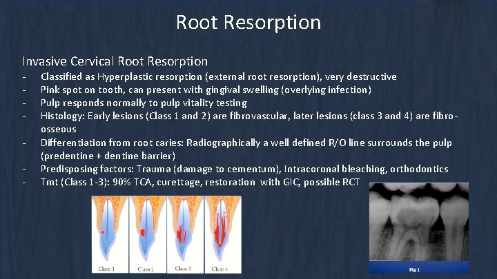
Root Resorption Invasive Cervical Root Resorption - Classified as Hyperplastic resorption (external root resorption), very destructive Pink spot on tooth, can present with gingival swelling (overlying infection) Pulp responds normally to pulp vitality testing Histology: Early lesions (Class 1 and 2) are fibrovascular, later lesions (class 3 and 4) are fibroosseous Differentiation from root caries: Radiographically a well defined R/O line surrounds the pulp (predentine + dentine barrier) Predisposing factors: Trauma (damage to cementum), Intracoronal bleaching, orthodontics Tmt (Class 1 -3): 90% TCA, curettage, restoration with GIC, possible RCT
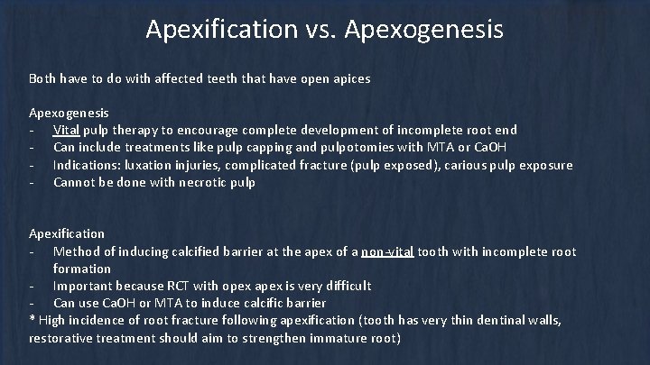
Apexification vs. Apexogenesis Both have to do with affected teeth that have open apices Apexogenesis - Vital pulp therapy to encourage complete development of incomplete root end - Can include treatments like pulp capping and pulpotomies with MTA or Ca. OH - Indications: luxation injuries, complicated fracture (pulp exposed), carious pulp exposure - Cannot be done with necrotic pulp Apexification - Method of inducing calcified barrier at the apex of a non-vital tooth with incomplete root formation - Important because RCT with opex apex is very difficult - Can use Ca. OH or MTA to induce calcific barrier * High incidence of root fracture following apexification (tooth has very thin dentinal walls, restorative treatment should aim to strengthen immature root)
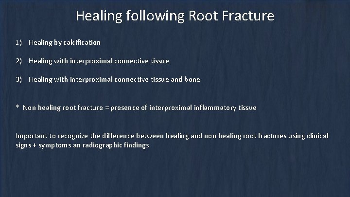
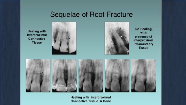
Healing following Root Fracture 1) Healing by calcification 2) Healing with interproximal connective tissue 3) Healing with interproximal connective tissue and bone * Non healing root fracture = presence of interproximal inflammatory tissue Important to recognize the difference between healing and non healing root fractures using clinical signs + symptoms an radiographic findings
Management of Trauma in Permanent Teeth Main Goal = Long term preservation of tooth if possible
Concussion DIAGNOSIS - Tooth is TTP, nil displacement or mobility - Pulp testing – usually +ve - Radiographically normal appearance TREATMENT - Not needed, monitor for 1 year - Clinical and radiographic control follow ups at: 4 weeks, 6 -8 weeks, 1 year PATIENT INSTRUCTIONS 1. Soft food: 7 days 2. Good OH: brush with a soft toothbrush after every meal, 0. 1% CHx gel topically bds 1/52 (if good toothbrushing not possible in the area)
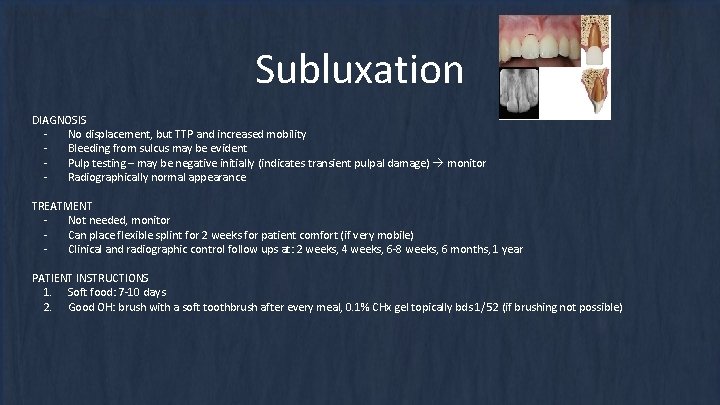
Subluxation DIAGNOSIS No displacement, but TTP and increased mobility Bleeding from sulcus may be evident Pulp testing – may be negative initially (indicates transient pulpal damage) monitor Radiographically normal appearance TREATMENT Not needed, monitor Can place flexible splint for 2 weeks for patient comfort (if very mobile) Clinical and radiographic control follow ups at: 2 weeks, 4 weeks, 6 -8 weeks, 6 months, 1 year PATIENT INSTRUCTIONS 1. Soft food: 7 -10 days 2. Good OH: brush with a soft toothbrush after every meal, 0. 1% CHx gel topically bds 1/52 (if brushing not possible)
Extrusion DIAGNOSIS Tooth appears elongated, TTP, and excessive mobility Pulp testing - unless minor, usually -ve. Radiographically increased PDL space apically TREATMENT 1. Reposition, LA usually not necessary 2. Stabilise for 2 weeks with flexible splint 3. Radiographically - open apex: can monitor, look for continued root development, +ve pulp testing. Closed apex: monitor 4. In closed apices where necrosis is anticipated, or where there are signs and symptoms consistent with pulpal necrosis RCT 5. Follow-up: 2 weeks, 4 weeks, 6 -8 weeks, 6 months, 1 yearly for 5 years PATIENT INSTRUCTIONS 1. Soft food: 7 -10 days 2. Good OH: brush with a soft toothbrush after every meal, 0. 1% CHx gel topically bds 1/52 (if brushing not possible) *Immature teeth: revascularisation usually occurs; mature teeth: may occur
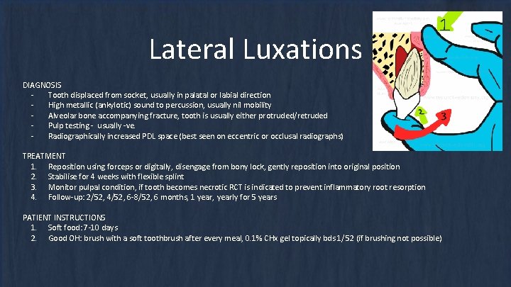
Lateral Luxations DIAGNOSIS Tooth displaced from socket, usually in palatal or labial direction High metallic (ankylotic) sound to percussion, usually nil mobility Alveolar bone accompanying fracture, tooth is usually either protruded/retruded Pulp testing - usually -ve. Radiographically increased PDL space (best seen on eccentric or occlusal radiographs) TREATMENT 1. Reposition using forceps or digitally, disengage from bony lock, gently reposition into original position 2. Stabilise for 4 weeks with flexible splint 3. Monitor pulpal condition, if tooth becomes necrotic RCT is indicated to prevent inflammatory root resorption 4. Follow-up: 2/52, 4/52, 6 -8/52, 6 months, 1 year, yearly for 5 years PATIENT INSTRUCTIONS 1. Soft food: 7 -10 days 2. Good OH: brush with a soft toothbrush after every meal, 0. 1% CHx gel topically bds 1/52 (if brushing not possible)
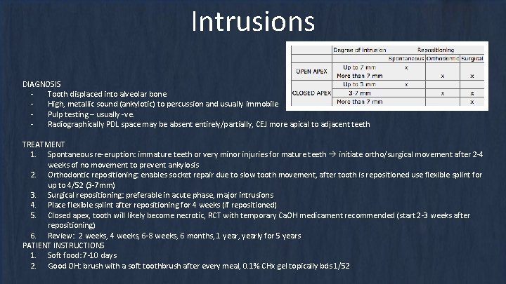
Intrusions DIAGNOSIS Tooth displaced into alveolar bone High, metallic sound (ankylotic) to percussion and usually immobile Pulp testing – usually -ve. Radiographically PDL space may be absent entirely/partially, CEJ more apical to adjacent teeth TREATMENT 1. Spontaneous re-eruption: immature teeth or very minor injuries for mature teeth initiate ortho/surgical movement after 2 -4 weeks of no movement to prevent ankylosis 2. Orthodontic repositioning: enables socket repair due to slow tooth movement, after tooth is repositioned use flexible splint for up to 4/52 (3 -7 mm) 3. Surgical repositioning: preferable in acute phase, major intrusions 4. Place flexible splint after repositioning for 4 weeks (if repositioned) 5. Closed apex, tooth will likely become necrotic, RCT with temporary Ca. OH medicament recommended (start 2 -3 weeks after repositioning) 6. Review: 2 weeks, 4 weeks, 6 -8 weeks, 6 months, 1 year, yearly for 5 years PATIENT INSTRUCTIONS 1. Soft food: 7 -10 days 2. Good OH: brush with a soft toothbrush after every meal, 0. 1% CHx gel topically bds 1/52
Avulsions At scene of the tragedy: - - Ensure it is a permanent tooth (primary teeth should not be replanted) Keep patient calm Pick up tooth by the crown, do not touch the root If tooth is dirty wash briefly (10 sec) under cold running water and reposition encourage patient/parent to reposition, bite on handkerchief to hold it in position If repositioning on site not possible store tooth in appropriate storage medium (goal = preserving viability of PDL cells) - HBSS Saline Saliva (I/O or E/O) AVOID storage in water Seek emergency dental treatment immediately
Avulsions (Closed Apex) Tooth replanted prior to arrival at clinic Treatment 1) Clean the area with water, saline, or CHx 2) Suture gingival lacerations 3) Verify normal position of tooth clinically + radiographically 4) Flexible splint for 2 weeks 5) Systemic antibiotics 7 days (tetracycline first choice, Pen V for children under 12) 6) If tooth was in contact with soil and tetanus status uncertain refer to GP for tetanus booster 7) RCT 7 -10 days (before removal of splint) use long term intra canal medicament (Ca. OH – 1 month, Odontopaste 2 weeks) 8) Review: 2 weeks, 4 weeks, 3 months, 6 months, 1 year, yearly thereafter (basically forever) Patient Instructions: - Avoid contact sports - Soft foods for up to 2 weeks - Brush teeth with soft toothbrush after each meal - Use 0. 1% CHx mouthrinse bds for 1 week
Avulsions (Closed Apex) E/O Dry time less than 60 mins Treatment 1) Clean root surface and apical foramen with stream of saline + soak tooth in saline (remove contamination + dead cells from root surface) 2) Give LA 3) Irrigate socket with saline 4) Examine socket for alveolar fracture, reposition fractured segment 5) Replant tooth with slight digital pressure 6) Suture gingival lacerations 6) Verify normal position of tooth clinically + radiographically 7) Flexible splint for 2 weeks 8) Systemic antibiotics 7 days (tetracycline first choice, Pen V for children under 12) 9) If tooth was in contact with soil and tetanus status uncertain refer to GP for tetanus booster 10) RCT 7 -10 days (before removal of splint) use long term intra canal medicament (Ca. OH – 1 month, Odontopaste 2 weeks) 11) Review: 2 weeks, 4 weeks, 3 months, 6 months, 1 year, yearly thereafter (basically forever) Patient Instructions: - Avoid contact sports - Soft foods for up to 2 weeks - Brush teeth with soft toothbrush after each meal - Use 0. 1% CHx mouthrinse bds for 1 week
Avulsions (Closed Apex) E/O Dry time more than 60 mins Treatment 1) Remove attached non-viable soft tissue carefully with gauze (can soak in 2% Na. F for 20 mins) 2) RCT can be done before replantation or 7 -10 days after replantation 3) Give LA *poor long term prognosis with this 4) Irrigate socket with saline scenario, goal is to maintain tooth 5) Examine socket for alveolar fracture, reposition fractured segment short term for aesthetics, function, and 6) Replant tooth with slight digital pressure psychological health + to maintain 7) Suture gingival lacerations bone contour however ankylosis and 8) Verify normal position of tooth clinically + radiographically root resorption is the expected 9) Flexible splint for 4 weeks eventual outcome 10) Systemic antibiotics 7 days (tetracycline first choice, Pen V for children under 12) 11) If tooth was in contact with soil and tetanus status uncertain refer to GP for tetanus booster 12) RCT 7 -10 days (or before replantation) use long term intra canal medicament (Ca. OH – 1 month, Odontopaste 2 weeks) 13) Review: 4 weeks, 3 months, 6 months, 1 year, yearly thereafter (basically forever) Patient Instructions: - Avoid contact sports - Soft foods for up to 2 weeks - Brush teeth with soft toothbrush after each meal - Use 0. 1% CHx mouthrinse bds for 1 week
Avulsions (Open Apex) Tooth replanted prior to arrival at clinic Signs of pulpal necrosis: - Negative response to pulp testing after at least 6 weeks - Development of classic signs re: PA R/L, TTP, draining sinus - Development of inflammatory root resorption (internal or external) Treatment 1) Clean the area with water, saline, or CHx 2) Suture gingival lacerations 3) Verify normal position of tooth clinically + radiographically 4) Flexible splint for 1 -2 weeks 5) Systemic antibiotics 7 days (tetracycline first choice, Pen V for children under 12) 6) If tooth was in contact with soil and tetanus status uncertain refer to GP for tetanus booster 7) Avoid RCT initially, if revascularisation fails and tooth becomes necrotic, perform RCT 8) Review: 2 weeks, 4 weeks, 3 months, 6 months, 1 year, yearly thereafter (basically forever) Patient Instructions: - Avoid contact sports - Soft foods for up to 2 weeks - Brush teeth with soft toothbrush after each meal - Use 0. 1% CHx mouthrinse bds for 1 week
Avulsions (Open Apex) E/O Dry time less than 60 mins Treatment 1) Clean root surface and apical foramen with stream of saline 2) Soak tooth in saline + antibiotics for 5 mins, 1 mg doxycycline per 20 ml saline (increases chances of revascularisation) 3) Give LA 4) Irrigate socket with saline 5) Examine socket for alveolar fracture, reposition fractured segment 6) Replant tooth with slight digital pressure 7) Suture gingival lacerations 8) Verify normal position of tooth clinically + radiographically 9) Flexible splint for 2 weeks 10) Systemic antibiotics 7 days (tetracycline first choice, Pen V for children under 12) 11) If tooth was in contact with soil and tetanus status uncertain refer to GP for tetanus booster 12) Avoid RCT initially, if revascularisation fails and tooth becomes necrotic, perform RCT 13) Review: 2 weeks, 4 weeks, 3 months, 6 months, 1 year, yearly thereafter (basically forever) Patient Instructions: - Avoid contact sports - Soft foods for up to 2 weeks - Brush teeth with soft toothbrush after each meal - Use 0. 1% CHx mouthrinse bds for 1 week
Avulsions (Open Apex) E/O Dry time more than 60 mins Treatment 1) Remove attached non-viable soft tissue carefully with gauze (can soak in 2% Na. F for 20 mins) 2) RCT can be done before replantation or 7 -10 days after replantation 3) Give LA No attempt made at revascularization 4) Irrigate socket with saline made here because of poor long term 5) Examine socket for alveolar fracture, reposition fractured segment prognosis, efforts made at delaying the 6) Replant tooth with slight digital pressure inevitable 7) Suture gingival lacerations 8) Verify normal position of tooth clinically + radiographically 9) Flexible splint for 4 weeks 10) Systemic antibiotics 7 days (tetracycline first choice, Pen V for children under 12) 11) If tooth was in contact with soil and tetanus status uncertain refer to GP for tetanus booster 12) RCT 7 -10 days (or before replantation) use long term intra canal medicament (Ca. OH – 1 month, Odontopaste 2 weeks) 13) Review: 4 weeks, 3 months, 6 months, 1 year, yearly thereafter (basically forever) Patient Instructions: - Avoid contact sports - Soft foods for up to 2 weeks - Brush teeth with soft toothbrush after each meal - Use 0. 1% CHx mouthrinse bds for 1 week
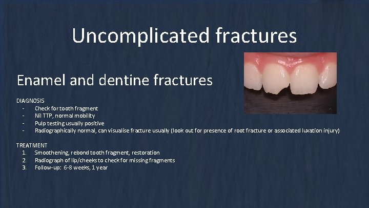
Uncomplicated fractures Enamel and dentine fractures DIAGNOSIS Check for tooth fragment Nil TTP, normal mobility Pulp testing usually positive Radiographically normal, can visualise fracture usually (look out for presence of root fracture or associated luxation injury) TREATMENT 1. Smoothening, rebond tooth fragment, restoration 2. Radiograph of lip/cheeks to check for missing fragments 3. Follow-up: 6 -8 weeks, 1 year
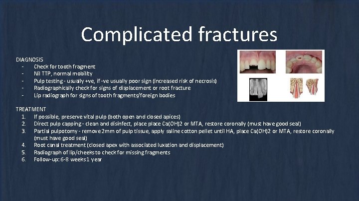
Complicated fractures DIAGNOSIS Check for tooth fragment Nil TTP, normal mobility Pulp testing - usually +ve, if -ve usually poor sign (increased risk of necrosis) Radiographically check for signs of displacement or root fracture Lip radiograph for signs of tooth fragments/foreign bodies TREATMENT 1. If possible, preserve vital pulp (both open and closed apices) 2. Direct pulp capping - clean and disinfect, place Ca(OH)2 or MTA, restore coronally (must have good seal) 3. Partial pulpotomy - remove 2 mm of pulp tissue, apply saline cotton pellet until HA, place Ca(OH)2 or MTA, restore coronally (must have good seal) 4. Root canal treatment (closed apex with associated luxation and displacement) 5. Radiograph of lip/cheeks to check for missing fragments 6. Follow-up: 6 -8 weeks 1 year
Subgingival fractures DIAGNOSIS Crown fracture extending subgingivally, may/may not involve pulp. Tooth is split into 2+ fragments, one of which is mobile Tenderness to percussion, at least one coronal fragment mobile, may have increased pain due to mobility Pulp testing - usually +ve for apical fragment Radiographically apical sign of fracture usually not visible TREATMENT 1. Fragment removal (small), restore coronally 2. Remove coronal segment, complete RCT, restore with post-core crown. Should be preceded with gingivectomy, ostectomy/osteoplasty. Only suitable with palatal subgingival extensions. 3. Orthodontic extrusion, placement of crown (implies RCT) 4. Surgical extrusion: remove mobile fragment, subsequently reposition root coronally, root rotation 90 to 180 degrees may offer better healing, extirpate and apply flexible splint. RCT can be commenced after 4 weeks. 5. Decoronation: postpones definitive restoration but preserves alveolar bone (implants) 6. Extraction 7. Follow-up: 6 -8 weeks, 1 year
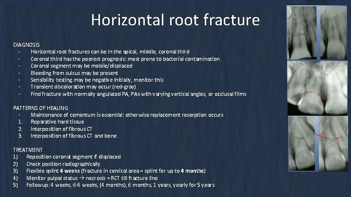
Horizontal root fracture DIAGNOSIS Horizontal root fractures can be in the apical, middle, coronal third Coronal third has the poorest prognosis: most prone to bacterial contamination Coronal segment may be mobile/displaced Bleeding from sulcus may be present Sensibility testing may be negative initially, monitor this Transient discoloration may occur (red-gray) Find fracture with normally angulated PA, PAs with varying vertical angles, or occlusal films PATTERNS OF HEALING Maintenance of cementum is essential: otherwise replacement resorption occurs 1. Reparative hard tissue 2. Interposition of fibrous CT 3. Interposition of fibrous CT and bone TREATMENT 1) Reposition coronal segment if displaced 2) Check position radiographically 3) Flexible splint 4 weeks (fracture in cervical area = splint for up to 4 months) 4) Monitor pulpal status necrosis = RCT till fracture line 5) Followup: 4 weeks, 6 -8 weeks, (4 months), 6 months, 1 years, yearly for 5 years
Alveolar fractures DIAGNOSIS - Displacement of alveolar segment, occlusal change due to misaligned segment - Tender to percussion, entire segment mobile, moves as unit - Pulp testing – may be negative - Occlusal change due to misalignment of fractured segment common - Radiographically check with 3 angulations, occlusal view, and OPG vertical line may run along PDL/septum, horizontal line may be located at any level. Gives valuable information about risk of damage to permanent tooth germs in children. TREATMENT 1. Manual repositioning under LA or use forceps (GA may be required in children) 2. Suture gingival lacerations 3. Apply flexible splint for 4 weeks 4. Follow-up: 4 weeks, 6 -8 weeks, 4 months, 6 months, 1 year, every year for 5 years PATIENT INSTRUCTIONS 1. Soft food: up to 2 weeks 2. Good OH: brush with a soft toothbrush after every meal, 0. 1% CHx gel topically bds 1/52
Management of Trauma in Primary Teeth Main Goal = To minimise damage to permanent successor tooth
Concussion DIAGNOSIS - Tooth is TTP, nil displacement or mobility - Radiographically normal appearance TREATMENT - Not needed - Clinical followup 1 week, 6 -8 weeks (nil radiographs required)
Subluxation DIAGNOSIS No displacement, but TTP and increased mobility Bleeding from sulcus may be evident Radiographically normal appearance Occlusal radiograph recommended check for displacement/root fracture, serves as baseline TREATMENT Not needed, monitor Clinical followup: 1 week, 6 -8 weeks OHI: Brush area with soft toothbrush + apply 0. 12% CHx gel with cotton bud 2/day for 1 week
Extrusion DIAGNOSIS Tooth partially displaced from socket Tooth appears elongated, mobility ++, Radiographically increased PDL space apically TREATMENT * Based on degree of displacement, mobility, root formation, and coping ability of child 1. If minor (<3 mm), immature developing tooth: careful reposition/leave for spontaneous realignment 2. If severe, fully formed permanent tooth = extraction 3. Follow-up: 1 week, 6/8 weeks, 6 months, 1 year (radiographs at 6/8 weeks, 6 months, 1 year) Tooth may become discoloured, monitor discoloured teeth for signs of infection (eg. draining sinus, loss of bone at furcation)
Lateral Luxation DIAGNOSIS Tooth displaced palatally or labially Nil mobility Occlusal radiograph: increased PDL space, position of displaced tooth in relation to permanent successor TREATMENT 1. No occlusal interference (eg. Anterior open bite) Spontaneous repositioning 2. Minor occlusal interference minor occlusal adjustment 3. More severe occlusal interference gentle repositioning under LA 4. Severe displacement extraction 5. Followup: 1 week, 2 -3 weeks, 6 -8 weeks, 1 year (radiographs at 6 -8 weeks, 1 year)
Intrusion DIAGNOSIS Tooth displaced through labial bone plate or can be impinging on permanent tooth bud Radiographs: if displaced through labial bone, can visualise apical tip + tooth appears shorter than contralateral, if impinging on permanent tooth bud the apical tip cannot be visualised and the tooth appears elongated TREATMENT 1. Apex displaced towards labial bone plate = spontaneous repositioning 2. Apex displaced towards permanent tooth germ = extract 3. Follow up: 1 week, 3 -4 weeks, 6 -8 weeks, 6 months, 1 year or until permanent tooth erupts (radiographs start at 3 -4 weeks)
Avulsion DIAGNOSIS Pretty straightforward TREATMENT 1. Do not replant primary teeth 2. Followup 1 week, 6 months, 1 year (or until permanent tooth erupts)
Uncomplicated Fracture DIAGNOSIS Fracture involving enamal and dentine, nil pulpal involvement TREATMENT 1. Smooth sharp edges, seal dentine with GIC, restore with CR 2. Clinical followup in 3 -4 weeks
Complicated Fracture DIAGNOSIS Check for tooth fragment Radiographically check for stage of root development TREATMENT 1. If possible, preserve vital pulp by partial pulpotomy (depends also on pt’s ability to cope) 2. Partial (Cvek) pulpotomy - remove 2 mm of pulp tissue, apply saline cotton pellet until HA, place Ca(OH)2 or MTA, restore coronally (must have good seal) 3. Otherwise Extraction 4. Radiograph of lip/cheeks to check for missing fragments 5. Follow-up: 1 week, 6 -8 weeks, 1 year
Subgingival fractures DIAGNOSIS Crown fracture extending subgingivally, may/may not involve pulp. Tooth is split into 2+ fragments, one of which is mobile Tenderness to percussion, at least one coronal fragment mobile, may have increased pain due to mobility Radiographically apical sign of fracture usually not visible, if lateral fracture check with relation to gingival margin TREATMENT 1. Depends on extent of fracture fragment removal or extraction 2. Pulpal involvement: partial pulpotomy, extraction 3. Followup: 1 weeks, 6 -8 weeks, 1 year
Horizontal root fracture (primary) DIAGNOSIS - Coronal segment usually mobile - Tooth possibly tender to percussion - PA or occlusal radiograph TREATMENT 1. If coronal segment not displaced = no treatment 2. If coronal segment mobile = extract and leave apical segment to be resorbed 3. Follow-up: 1/52, 6 -8/52, 1 year PATIENT INSTRUCTIONS 1. Soft food: 10 -14 days 2. Good OH: brush with a soft toothbrush after every meal, 0. 1% CHx gel topically bds 1/52 3. Monitor for signs of pulpal pathosis
Alveolar Fractures (Children) DIAGNOSIS - Fractures that involve alveolar bone may involve adjacent bones - Segment mobility/dislocation + occlusal interferences common TREATMENT 1. GA often indicated 2. Reposition displaced segment, splint for 4 weeks 3. Monitor teeth associated with fracture 4. Followup: 1 week, 3 -4 weeks, 6 -8 weeks, 1 year (or until permanent tooth erupts)
Questions?