Throat and Tongue 2 Muscles grouped for function

- Slides: 16
Throat and Tongue 2
Muscles grouped for function • • Facial Expression Eye movements Move the lower jaw Soft palate Pharynx Larynx The tongue – Extrinsic – move the tongue – Intrinsic – inside the tongue
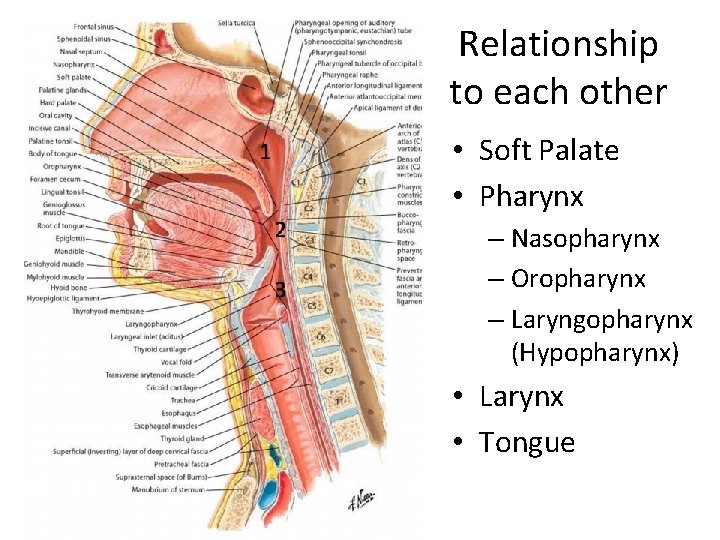
Relationship to each other • Soft Palate • Pharynx – Nasopharynx – Oropharynx – Laryngopharynx (Hypopharynx) • Larynx • Tongue
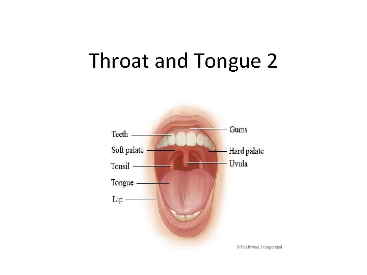
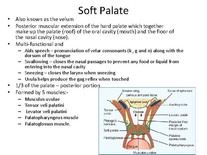
Soft Palate • Also known as the velum • Posterior muscular extension of the hard palate which together make up the palate (roof) of the oral cavity (mouth) and the floor of the nasal cavity (nose). • Multi-functional and – Aids speech – pronunciation of velar consonants (k , g and n) along with the dorsum of the tongue – Swallowing – closes the nasal passages to prevent any food or liquid from entering into the nasal cavity – Sneezing – closes the larynx when sneezing – Uvula helps produce the gag reflex when touched • 1/3 of the palate – posterior portion • Formed by 5 muscles: – – – Musculus uvulae Tensor veli palatini Levator veli palatini Palatopharyngeus muscle Palatoglossus muscle.
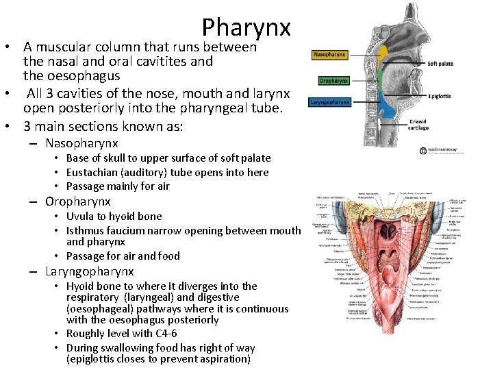
Pharynx • A muscular column that runs between the nasal and oral cavitites and the oesophagus • All 3 cavities of the nose, mouth and larynx open posteriorly into the pharyngeal tube. • 3 main sections known as: – Nasopharynx • Base of skull to upper surface of soft palate • Eustachian (auditory) tube opens into here • Passage mainly for air – Oropharynx • Uvula to hyoid bone • Isthmus faucium narrow opening between mouth and pharynx • Passage for air and food – Laryngopharynx • Hyoid bone to where it diverges into the respiratory (laryngeal) and digestive (oesophageal) pathways where it is continuous with the oesophagus posteriorly • Roughly level with C 4 -6 • During swallowing food has right of way (epiglottis closes to prevent aspiration)
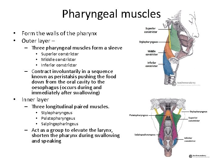
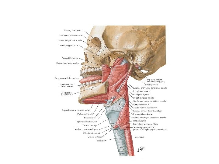
Pharyngeal muscles • Form the walls of the pharynx • Outer layer – – Three pharyngeal muscles form a sleeve • Superior constrictor • Middle constrictor • Inferior constrictor – Contract involuntarily in a sequence known as peristalsis pushing the food down from the oral cavity to the oesophagus (occurs during and immediately after swallowing) • Inner layer – Three longitudinal paired muscles. • Stylopharyngeus • Palatopharyngeus • Salpingopharingeus – Act as a group to elevate the larynx, shorten the pharynx during swallowing and speaking
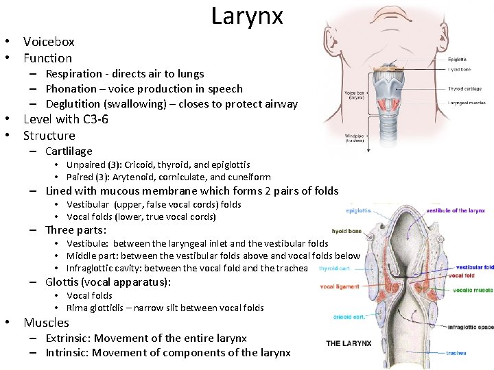
Larynx • Voicebox • Function – Respiration - directs air to lungs – Phonation – voice production in speech – Deglutition (swallowing) – closes to protect airway • Level with C 3 -6 • Structure – Cartlilage • Unpaired (3): Cricoid, thyroid, and epiglottis • Paired (3): Arytenoid, corniculate, and cuneiform – Lined with mucous membrane which forms 2 pairs of folds • Vestibular (upper, false vocal cords) folds • Vocal folds (lower, true vocal cords) – Three parts: • Vestibule: between the laryngeal inlet and the vestibular folds • Middle part: between the vestibular folds above and vocal folds below • Infraglottic cavity: between the vocal fold and the trachea – Glottis (vocal apparatus): • Vocal folds • Rima glottidis – narrow slit between vocal folds • Muscles – Extrinsic: Movement of the entire larynx – Intrinsic: Movement of components of the larynx
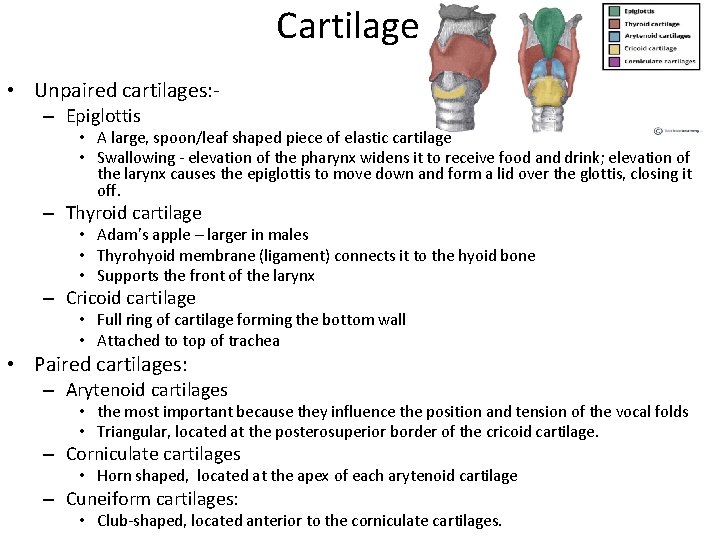
Cartilage • Unpaired cartilages: – Epiglottis • A large, spoon/leaf shaped piece of elastic cartilage • Swallowing - elevation of the pharynx widens it to receive food and drink; elevation of the larynx causes the epiglottis to move down and form a lid over the glottis, closing it off. – Thyroid cartilage • Adam’s apple – larger in males • Thyrohyoid membrane (ligament) connects it to the hyoid bone • Supports the front of the larynx – Cricoid cartilage • Full ring of cartilage forming the bottom wall • Attached to top of trachea • Paired cartilages: – Arytenoid cartilages • the most important because they influence the position and tension of the vocal folds • Triangular, located at the posterosuperior border of the cricoid cartilage. – Corniculate cartilages • Horn shaped, located at the apex of each arytenoid cartilage – Cuneiform cartilages: • Club-shaped, located anterior to the corniculate cartilages.
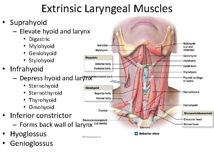
Extrinsic Laryngeal Muscles • Suprahyoid – Elevate hyoid and larynx • • Digastric Mylohyoid Geniohyoid Stylohyoid • Infrahyoid – Depress hyoid and larynx • • Sternohyoid Sternothyroid Thyrohyoid Omohyoid • Inferior constrictor – Forms back wall of larynx • Hyoglossus • Genioglossus
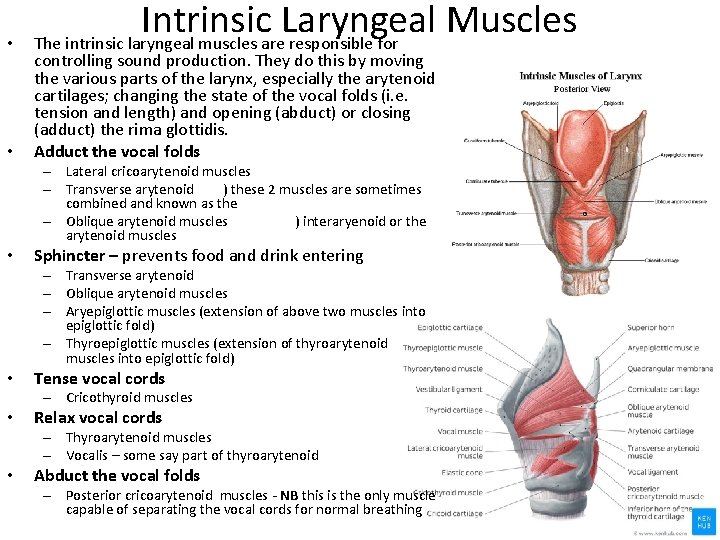
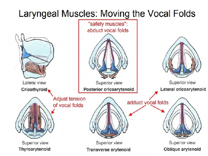
• • Intrinsic Laryngeal Muscles The intrinsic laryngeal muscles are responsible for controlling sound production. They do this by moving the various parts of the larynx, especially the arytenoid cartilages; changing the state of the vocal folds (i. e. tension and length) and opening (abduct) or closing (adduct) the rima glottidis. Adduct the vocal folds – Lateral cricoarytenoid muscles – Transverse arytenoid ) these 2 muscles are sometimes combined and known as the – Oblique arytenoid muscles ) interaryenoid or the arytenoid muscles • Sphincter – prevents food and drink entering – Transverse arytenoid – Oblique arytenoid muscles – Aryepiglottic muscles (extension of above two muscles into epiglottic fold) – Thyroepiglottic muscles (extension of thyroarytenoid muscles into epiglottic fold) • Tense vocal cords – Cricothyroid muscles • Relax vocal cords – Thyroarytenoid muscles – Vocalis – some say part of thyroarytenoid • Abduct the vocal folds – Posterior cricoarytenoid muscles - NB this is the only muscle capable of separating the vocal cords for normal breathing
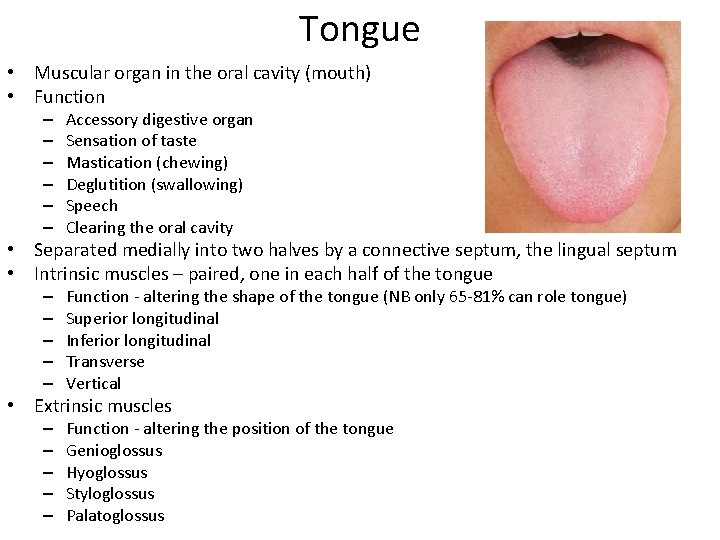
Tongue • Muscular organ in the oral cavity (mouth) • Function – – – Accessory digestive organ Sensation of taste Mastication (chewing) Deglutition (swallowing) Speech Clearing the oral cavity • Separated medially into two halves by a connective septum, the lingual septum • Intrinsic muscles – paired, one in each half of the tongue – – – Function - altering the shape of the tongue (NB only 65 -81% can role tongue) Superior longitudinal Inferior longitudinal Transverse Vertical • Extrinsic muscles – – – Function - altering the position of the tongue Genioglossus Hyoglossus Styloglossus Palatoglossus
Tongue muscles Work together to produce movements that are essential for mastication (chewing), all speech, and deglutition (swallowing). Intrinsic • Not attached to bone • Direction of fibres give the name • Change shape • Actions – Lengthen – Shorten – Curl/uncurling its apex and edges (tongue rolling) – Flatten/round surface • Muscles – – Superior longitudinal Inferior longitudinal Transverse Vertical • • Extrinsic Anchored to bone Bony origins give the name Change position Actions – Protrusion – Retraction – Side to side • Muscles – – Genioglossus Styloglossuss Palatoglossus Hyoglossus
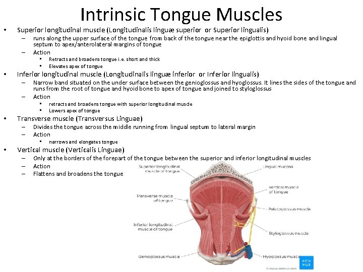
• Intrinsic Tongue Muscles Superior longitudinal muscle (Longitudinalis linguæ superior or Superior lingualis) – – runs along the upper surface of the tongue from back of the tongue near the epiglottis and hyoid bone and lingual septum to apex/anterolateral margins of tongue Action • • • Inferior longitudinal muscle (Longitudinalis linguæ inferior or Inferior lingualis) – – Narrow band situated on the under surface between the genioglossus and hyoglossus. It lines the sides of the tongue and runs from the root of tongue and hyoid bone to apex of tongue and joined to styloglossus Action • • • retracts and broadens tongue with superior longitudinal muscle Lowers apex of tongue Transverse muscle (Transversus Linguae) – – Divides the tongue across the middle running from lingual septum to lateral margin Action • • Retracts and broadens tongue i. e. short and thick Elevates apex of tongue narrows and elongates tongue Vertical muscle (Verticalis Linguae) – – – Only at the borders of the forepart of the tongue between the superior and inferior longitudinal muscles Action Flattens and broadens the tongue
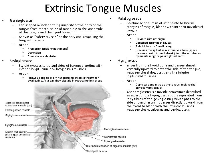
Extrinsic Tongue Muscles • Genioglossus – – – Protrusion (sticking out tongue) Depresion Contralateral deviation Styloglossuss – – Styloid process to tip and sides of tongue blending with inferior longitudinal and hyoglossus muscles Action • draws up the sides of the tongue to create a trough for swallowing. As a pair they also aid in retracting the tongue Palatoglossus – Fan shaped muscle forming majority of the body of the tongue from mental spine of mandible to the underside of the tongue and the hyoid bone Known as “safety muscle” as the only one propelling the tongue forwards Action • • • – palatine aponeurosis of soft palate to lateral margins of tongue, blends with intrinsic muscles of tongue Action • • • Elevates root of tongue Constricts isthmus of fauces Aids initiation of swallowing Prevents the spill of saliva from vestibule (space between teeth lips and cheeks) into the oropharynx by maintaining the palatoglossal arch. Hyoglossus – – arises from the hyoid bone and passes almost vertically upward to enter the side of the tongue, between the styloglossus and the inferior logitudinal muscles Action • – Depresses and retracts the tongue, making the surface more convex Chondroglossus is a muscle sometimes described as a part of the hypoglossus but is separated from it by fibres of the genioglossus, which pass to the side of the pharynx. It passes directly upward from the hyoid to blend with the intrinsic muscles between the hyoglossus and genioglossus