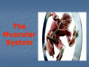
THE MUSCULAR SYSTEM The muscular system moves the


- Slides: 29

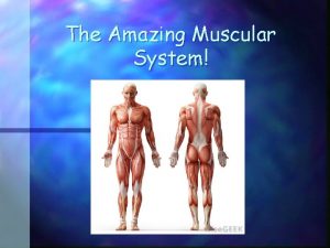
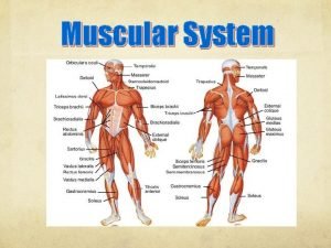
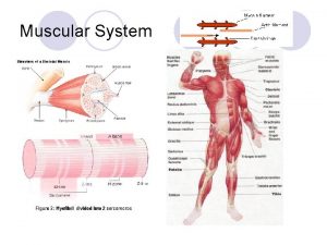
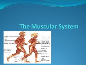
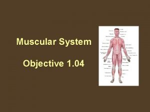
THE MUSCULAR SYSTEM The muscular system moves the body, maintains posture and produces heat. There are over 600 skeletal muscles which enable the human body to move. And muscles make up approximately 40 percent of the body’s weight.

Some of the Skeletal Muscles of the Skeletal. Muscular System in Humans

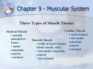
Types of muscles: Skeletal, Cardiac and Smooth. � � The skeletal muscles attached to bones allow for voluntary movement. Other muscles such as the muscles involved in focusing of the eyes work without us having to think about them working. Unlike cardiac and smooth muscle cells, skeletal muscles have many nuclei because they are formed from the fusion of many cells. Cardiac muscles found only in the walls of the heart, contract and relax continuously as they pump blood throughout the body. Smooth muscles in the throat, force food down into the esophagus, smooth muscles in the bladder squeeze urine out, and smooth muscles in the uterine wall push the baby out of the womb at childbirth. The skeletal muscles that function with the bones to allow for movement will be the focus of this chapter. The other muscle types were introduced in the chapter on tissue and will be covered in other chapters.

Interactions Between Muscles and Bones � � � Skeletal muscles contain bundles of hundreds of muscle cells which look like long striped fibers and are attached to bones. Muscles contract (shorten) in response to stimulation. In addition to many other activities muscles are necessary for humans to swallow, breathe, run, and even to write. Tendons attach muscles to bones. The tendons are made up of dense connective tissues. These connective tissues package muscles cells together and then extend as tendons. Muscles connect to bones near joints. Muscles contract and apply force making bones move. The tendons rub against bones and the synovial fluid filled cavities reduce friction.

Muscle Contraction and Movement. � � � The skeletal muscles work together in pairs or groups and enable movement. Some muscles work in opposition whereby the action of one opposes the action of another. For example, the biceps and triceps are connected to bones in the shoulder and extend to bones in the forearm. A muscle can only move in one direction and pulls the bones to which it connects as it contracts. A different muscle is needed to reverse the action. The biceps and triceps are examples of muscles working in opposition. The bicep muscles contract and raise the forearm and the elbow bends as we raise for instance, an apple in our hands to our mouths. Lowering the forearm after taking a bite of the apple requires the triceps to contract pulling the forearm bone down.

Opposing muscle groups: the triceps relaxed, the biceps contracted and vice versa.

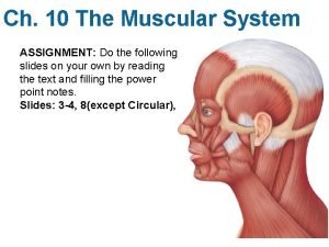
Muscle Fibers and How They Contract. � � � Some of the skeletal muscles of humans are superficial while others are deep in the walls of the body. Some of the facial muscles are attached to the skin. The trunk of the body has muscles of the thorax, backbone, abdominal wall and the pelvic cavity. Other muscles attach to limbs. Muscles are made of long thin cells known as fibers that are organized by the hundreds into groups called fascicles. Muscles are shaped like elongated cylinders with all the cells running parallel. The amount of fibers present is determined by the function the muscle will perform. Fibers can be white, contracting readily for actions that require force and power, or red performing slow contractions in movements of force and sustained traction.

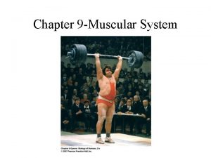
White Muscle and Red Muscle Fibers. � White muscle fibers also called fast-twitch fibers have fewer mitochondria, little or no myoglobin, and fewer blood vessels. The myosin in fast-twitch muscles has high ATP enzyme activity that catalyzes the breakdown of ATP. � These fibers are good for work that require maximum strength for a short period of time. Sprinters, and weight lifters usually have a high concentration of fast-twitch fibers in their leg and arm muscles. � Red muscle fibers also known as slow-twitch muscle fibers have a lot of the oxygen binding molecule myoglobin, a lot of mitochondria, and a lot of blood vessels. These fibers produce low tension and develop slowly but they can provide the energy needed to perform for an extended period. � The red muscle fibers have a large reserve of fuel (glycogen and fat) so if oxygen is available these fibers can continue to produce ATP for a few hours. Long distant runners, and cyclists have leg and arm muscles with mostly slow-twitch fibers.

Performance Capabilities of Different Muscle Fibers. � � � Aerobic training, can to some extent, alter the type of muscle fiber present in an individual. However, genetic inheritance is the key player in determining whether a person is a champion sprinter or a cross country runner. The amount of mitochondria in fast-twitch muscle fibers can be improved with aerobic exercises thus allowing for prolonging activities. Likewise a person with slow–twitch muscle fiber can train to increase speed. All human muscles have both fast-twitch and slow-twitch muscle fibers but in different percentages. Marathon runners like the 2016 Olympic winner, Jemima Jelagat Sumgong may have as much as 80 percent slow-twitch (red muscle) fiber in their muscle. Champion sprinters, like the eight time Olympic gold medalist Usain Bolt have about 60 percent fast-twitch (white muscle) fibers.

Muscle Fibers and How They Contract. � � � Each muscle has in its structure numerous, fine, thread-like material, (filament) known as myofibrils. The myofibrils have two main classes of proteins, myosin (thick filaments) and actin thinner filament. These two kinds of fibers, myosin and actin are arranged in matrices (matrixsingular) called sarcomeres. (Other proteins are also found in the sarcomeres. ) Skeletal muscles are described as striated because of the repeating units of sarcomeres along the length of the myofibril which when stained and observed under the light microscope appear striped. The structural arrangement of the sarcomere causing the striped appearance has alternating bands of actin molecule, the thin filament, and myosin molecules, the thick filament.

The Structure of the Skeletal Muscle

Organization and Function of the Muscle Fiber. � � The sarcomere is the contractile region in the myofibril, the unit of action. The arrangement of the thick and thin filament is the key to how sarcomeres, and the whole muscle contracts. The thin filament each made of two tightly coiled strands of actin monomers have tropomyosin, and troponin and other regulatory proteins bound tightly along its length. The thick filament, myosin molecules are motor proteins with club-like shaped head region. Each sarcomere is bound by what are called Z lines, which are structures that hold the thin actin filaments in place. (Z lines are made of a mesh of cytoskeleton elements). Z lines mark the boundary between sarcomeres and keep them anchored. Proteins called desmins and other intermediate filaments around each Z line help keep neighboring sarcomeres aligned.

Organization and Function of the Muscle Fiber continued. � � � Each sarcomere is made of overlapping filaments of actin and myosin. When the muscles contract, the sarcomeres shorten and the appearance of the band pattern changes. As stated before, each sarcomere is bound by Z lines, structures anchoring the thin actin. In the center of the sarcomeres are A bands containing all the myosin filaments. H zones and the I bands, light in color, are the areas where the actin and myosin do not overlap in the relaxed muscle. The dark striped area within the H zones is called the M band contains proteins that help hold the myosin filaments in their regular hexagonal arrangement.

Organization and Function continued. � � An elastic filament protein called titin is also anchored to Z bands and runs parallel to the rest. These titins keep the myosin centered in a contracting sarcomere and is responsible for the muscle not stretching while relaxed. All parts of the muscle bundle; muscle cells, myofibrils, thick, thin, and elastic filaments all have the same parallel orientation. All parts must maintain the parallel orientation to ensure that the force of contraction on a bone is only in one direction. The sliding-filament model is what has been accepted by scientists to explain muscle contractions.

The Sliding-filament Model of Muscle Contraction. � � � When the muscle contracts the width of the sarcomere shortens when its thin filaments are moved along by its thick filament. All the myosin filaments remain stationary and energy obtained from ATP is used to slide the two sets of actin filament over the myosin filament towards the center of the sarcomere. Both sets of actin slide over the myosin. The head of the myosin, the club-like shaped region, binds repeatedly to sites on the actin filament. The head of the myosin is an enzyme that hydrolyzes ATP. Each binding between the myosin and actin is called a cross- bridge. The myosin head is only able to form the cross-bridge after local concentration of calcium ions expose a binding site.

The Sliding-filament Model for Muscle Contraction continued. � � � The formation of the cross-bridge causes the head of the myosin to pull the actin to the center of the sarcomere. Next ATP then binds to the head of the myosin and causes the myosin to lose its grip on the actin. The ATP becomes hydrolyzed and the energy released makes the head fall back to its original position. The concentration of calcium determines whether or not the myosin head attaches to another binding site. A single contraction involves hundreds of myosin filaments moving down the entire length of the actin filament.

The Mechanism of Filament Sliding

The Role of the Nervous System in Muscular Contractions. � � � Skeletal muscles are under the control of the nervous system. The commands are relayed by motor neurons and stimulate or inhibit the sarcomeres. Muscle cells like all others have different electrical charge across the membranes. The cytoplasm just below the membrane is a bit more negative than the interstitial fluid surrounding it on the outside. Muscle cells, neurons, and other excitable cells can have a brief and abrupt reversal in the difference in the charge. This brief abrupt reversal of charges across the membrane is known as an action potential. It happens when charged ions cross the membranes in an accelerated rate. Such disturbance spreads away from the stimulation point without diminishing in intensity.

Motor Neuron and the Muscle Fiber Being Controlled.

The Sarcoplasmic Reticulum � � � When an action potential arises in a muscle cell, it spreads rapidly away from the stimulation point, and spreads along tubular extensions of the plasma membrane. These tubular extensions extend into and branch throughout the cytoplasm also known as the sarcoplasm of muscle fiber. The action potential that spreads across the plasma membrane also reaches these tubular extensions known as transverse tubules or T tubules. There anchor points on the T tubules for the actin filament. T tubules also connect with a system of membranous chambers that wrap around the myofibril. The system called sarcoplasmic reticulum has calcium pumps that cause membrane enclosed compartments of the cell to take in calcium ions thus acting as a storage site for calcium ions.

The Role of Calcium Ions in Muscle Contraction. � � � The arrival of the action potential causes calcium ions to flow from these membranous chambers. The released ions diffuse into myofibrils and reach actin. In resting muscle cells, the actin binding sites are blocked and myosin cannot form cross-bridge. The released calcium ions clear the binding sites so contraction can proceed. After contraction, calcium ions are actively transported back into sarcoplasmic reticulum. Video Link: Skeletal Muscle Contraction

T Tubules in Action

The Specific Role of the Proteins Tropomyosin and Troponin. � � � Tropomyosin filaments and troponins are located in or near the grooves on the surface of actin filament. When calcium level is low the two proteins are joined tightly that the tropomyosin is forced slightly outside the groove. It therefore blocks the myosin cross-bridge binding site. When there is a signal from the nervous system for the muscle to contract, calcium ions are released from the sarcoplasmic reticulum and flow into the sarcomere. The calcium ions bind with the troponin and change its shape. This changes the grip the troponin has on the tropomyosin filament and the tropomyosin filament is now free to move into the groove, exposing the binding site on the acting filament so the cross-bridge can be formed.

Energy for Contraction. � � � Cells need energy but muscles cells have a greater demand. When muscle cells contract the demand for phosphate from ATP must occur twenty to a hundred times faster than with other cells. However, only a small amount of ATP is available and therefore enzymes transfer the phosphate from creatine phosphate (phosphocreatine CP or PCr) an organic compound, to ADP. Cells have five times as much creatine phosphate as ATP and so the process of energy production can be maintained until the slower ATP formation starts.

Anaerobic Down of Glucose in Fast-twitch Muscle Fiber. � � � Sometimes the muscle cells undergo glycolysis, the anaerobic breakdown of glucose to provide small amounts of ATP. The glucose is not completely broken down. Lactic acid forms as the end-product instead of carbon dioxide. The lactic acid causes muscle aches and fatigue. This lactic acid does not remain in the muscles but is transported in the blood to the liver where it is converted back to pyruvic acid.

Muscle Tension � � � The external force acting on a muscle will determine if the muscle actually contracts. Cross-bridge exerts muscle tension. Muscle tension is the mechanical force that a contracting muscle exerts on an object like a bone. Opposing the tension is a load and the muscle will only contract if the muscle tension exceeds the load. Isotonically contracting muscles shorten and move a load. An isometrically contracting muscle develops tension but does not move a load.

Muscle Fatigue and Muscular Dystrophies. � � Muscle fatigue occurs when the muscle is not able to generate force. This can happen when the muscle is overworked from intense exercise like weigh lifting. Resting the muscle will correct the problem. There are different types of muscular dystrophies. These are genetic disorders. The diseases are X-linked. The mutation results in decreased amount or complete lack of the protein dystrophin. Dystrophin is needed to bond to actin in order for the sarcomere to contract. Duchenne dystrophy is the most common form in children Myotonic muscular dystrophy is the most common form in adults. Muscular dystrophy affects mainly the skeletal muscles.

Muscular Performance and Aging. � � � Muscle tension declines with age. With activity muscle cells increase in size, muscle activity and become resistant to fatigue. Aerobic exercise causes an increase in mitochondria and blood capillaries. More blood capillaries mean more glucose and oxygen can be brought to the muscles and more waste can be gotten rid of efficiently. Increased amounts of mitochondria result in the more efficient break down of glucose and more oxygen carried to the muscles by the increased blood vessels. As we age muscle tension declines and so the need for continued exercise is vital. Strength training is important as it slows the loss of muscle.

Usain Bolt Record Breaking Races at the Beijing Olympics in 2008 � Usain Bolt at Beijing 2008
 Differentiate muscular strength from muscular endurance
 A storm system moves 5000 km due east
 A storm system moves 5000 km due east
 700 calories scalar or vector
 A storm system moves 5000 km due east
 A storm system moves 5000 km due east
 A bear searching for food wanders 35 meters east
 Three functions of muscular system
 Whats the function of the muscular system
 Function of skeletal muscle
 Chapter 4 the muscular system learning exercises answer key
 Chapter 6 the muscular system figure 6-9
 Simple muscular system diagram
 Endomysium
 Muscular system label
 Rectus femoris fascicle arrangement
 Muscular system head and neck
 Sheep muscular system
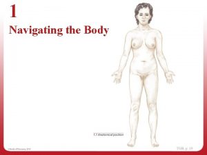
 Navigating the body
 Whats the function of the muscular system
 Chapter 6 the muscular system figure 6-12
 Whats muscular system
 Chapter 7:5 muscular system
 Chapter 9 muscular system
 Structures of the muscular system
 Active muscle
 Chapter 32 section 3 the muscular system answer key
 Muscular system response to exercise
 Whats the muscular system
 Muscular system diagram

