The Eye Extrinsic Eye Muscles Six skeletal muscles


- Slides: 15
The Eye
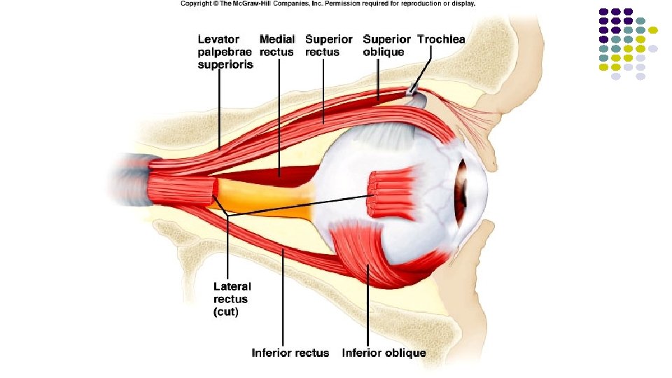
Extrinsic Eye Muscles Six skeletal muscles that move the eyeball ● ● ● Superior rectus- rotates eye up and medially Inferior rectus- rotates eye down and medially Medial rectus- rotates eye medially Lateral rectus- rotates eye laterally Superior oblique- rotates eye down and laterally Inferior oblique- rotates eye up and laterally
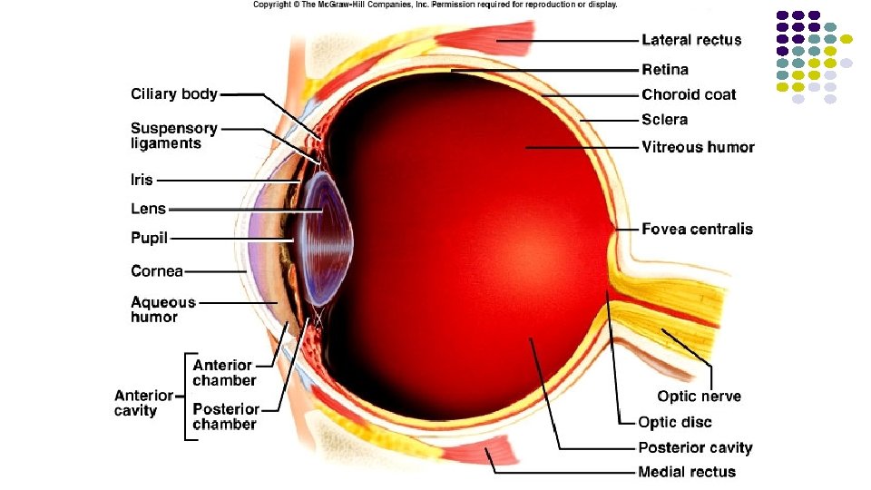
Eye Structure and Function 3 layers: 1. Outer Fibrous Tunic 2. Middle Vascular Tunic 3. Inner Nervous Tunic
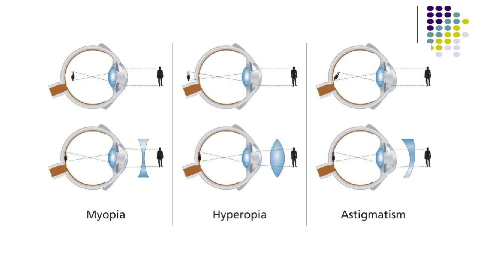
1. Outer Fibrous Tunic Cornea- anterior portion transparent, light transmission, light refraction ● Sclera- posterior portion, opaque, protection ● Lens-Transparent, lies behind iris, largely composed of lens fibers, elastic, By changing its shape, the lens changes the focal distance of the eye. In other words, it focuses the light rays that pass through it (and onto the retina) in order to create clear images of objects that are positioned at various distances. ●
2. Middle Vascular Tunic ● ● ● Iris- anterior, pigmented, controls light intensity by adjusting the size of the pupil – the opening in the middle of the iris Ciliary body- anterior, pigmented, holds lens, moves lens for focusing Choroid coat- provides blood supply, pigments absorb extra light ● ● Tapetum lucidum –a layer of tissue in the eye of many vertebrates. Lying immediately behind the retina, it is a retroreflector. Contributes to the superior night vision of some animals. Anterior chamber of eye filled with a watery liquid called aqueous humor.
3. Inner Nervous Tunic ● retina ▪ contains visual receptors ▪ continuous with optic nerve ▪ ends just behind margin of the ciliary body ▪ composed of several layers ● fovea centralis –produces sharpest vision ● optic disc – the raised disk on the retina at the point of entry of the optic nerve, lacking visual receptors and so creating a blind spot. ● vitreous humor – thick gel that holds retina flat against choroid coat
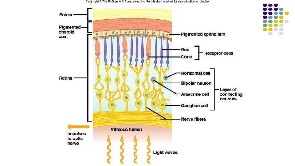
How We See ● ● Photoreceptors in the retina: Rods- long, thin projections, contain light sensitive pigment called rhodopsin, hundred times more sensitive to light than cones, provide vision in dim light, produce colorless vision, produce outlines of objects
How We See ● ● Photoreceptors in the retina: Cones- short, blunt projections, contain light sensitive pigments called erythrolabe(Red Light), chlorolabe(Green Light), and cyanolabe(Blue Light), provide vision in bright light, produce sharp images, produce color vision
How We See ● Fovea centralis: a tiny pit located in the macula of the retina that provides the clearest vision of all. Only here the layers of the retina spread aside to let light fall directly on the cones, the cells that give the sharpest image.
How we see ● https: //slideplayer. com/slide/6905038/