Radiological manifestations of common MSK conditions Dr Richard

- Slides: 15
Radiological manifestations of common MSK conditions Dr Richard Beese Dr Sheela Anpalakhan Online webinar 18/7/2018
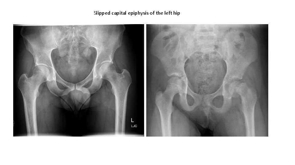
Slipped capital epiphysis of the left hip
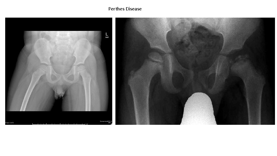
Perthes Disease
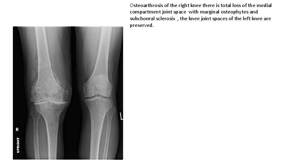
Osteoarthrosis of the right knee there is total loss of the medial compartment joint space with marginal osteophytes and subchonral sclerosis , the knee joint spaces of the left knee are preserved.
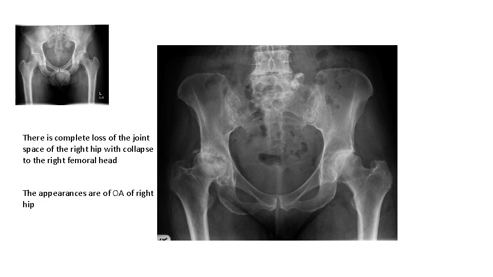
There is complete loss of the joint space of the right hip with collapse to the right femoral head The appearances are of OA of right hip
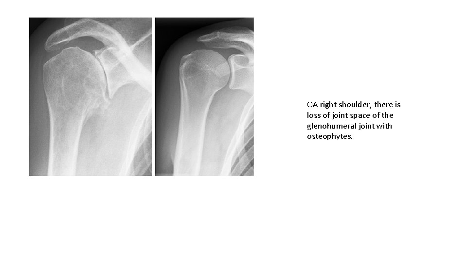
OA right shoulder, there is loss of joint space of the glenohumeral joint with osteophytes.
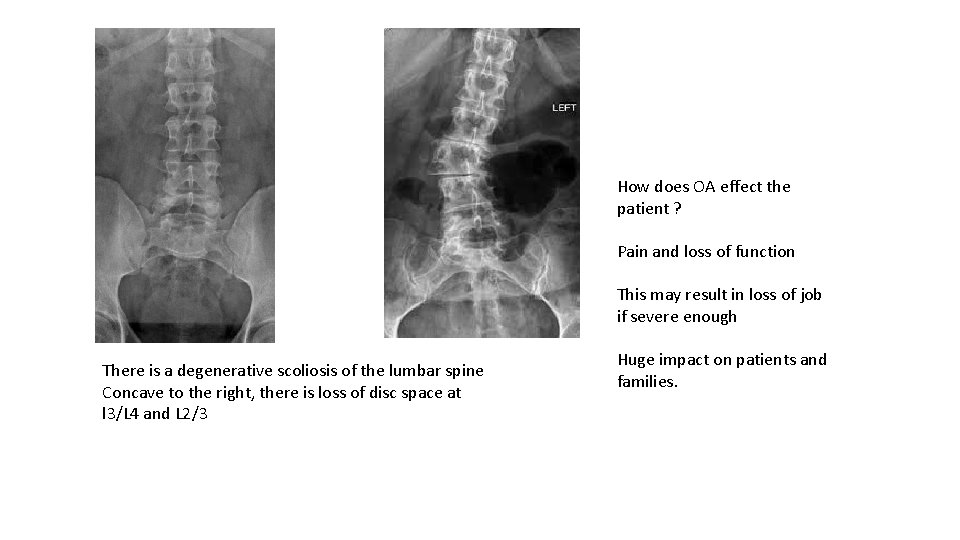
How does OA effect the patient ? Pain and loss of function This may result in loss of job if severe enough There is a degenerative scoliosis of the lumbar spine Concave to the right, there is loss of disc space at l 3/L 4 and L 2/3 Huge impact on patients and families.
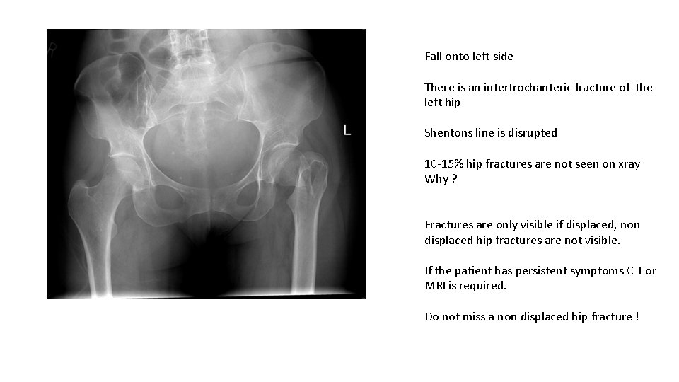
Fall onto left side There is an intertrochanteric fracture of the left hip Shentons line is disrupted 10 -15% hip fractures are not seen on xray Why ? Fractures are only visible if displaced, non displaced hip fractures are not visible. If the patient has persistent symptoms C T or MRI is required. Do not miss a non displaced hip fracture !
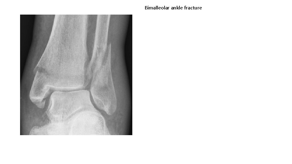
Bimalleolar ankle fracture
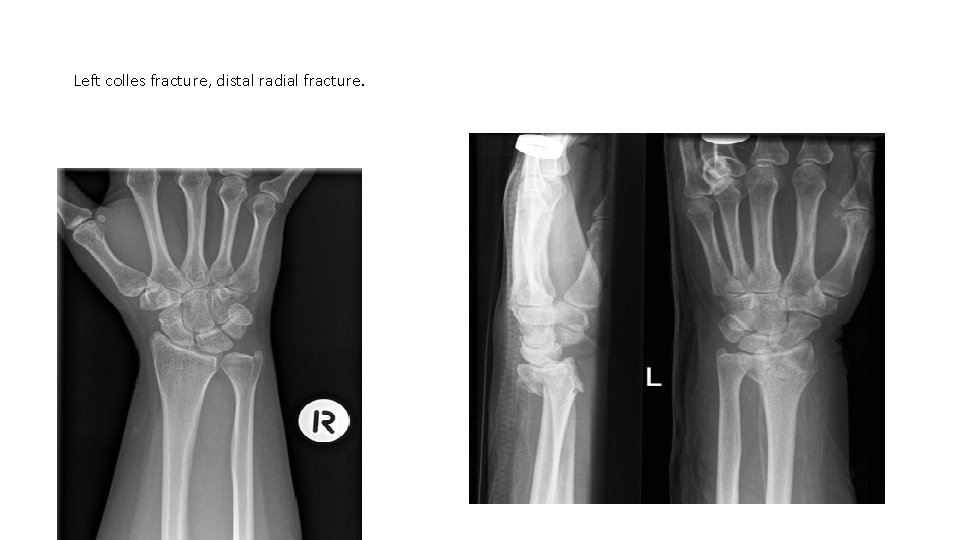
Left colles fracture, distal radial fracture.
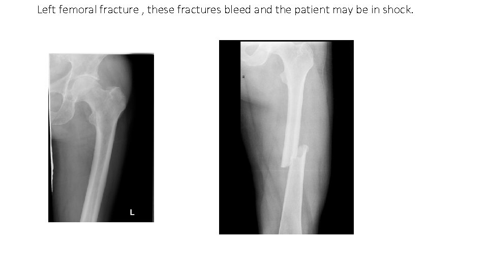
Left femoral fracture , these fractures bleed and the patient may be in shock.
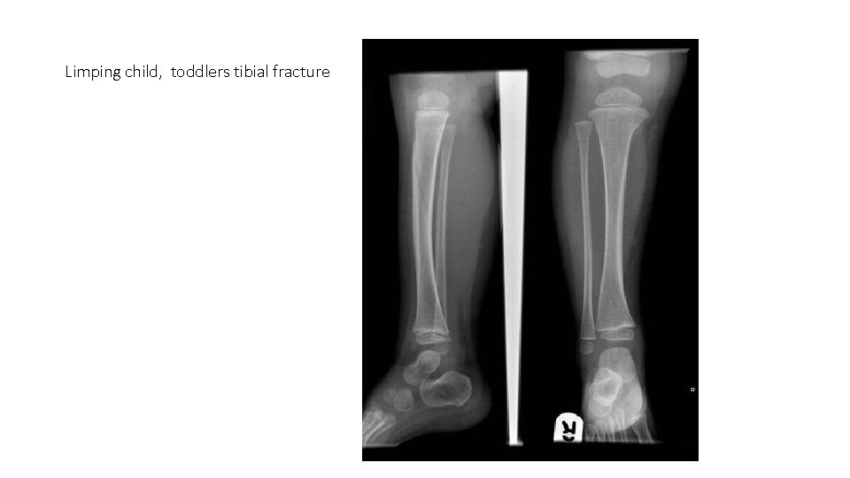
Limping child, toddlers tibial fracture
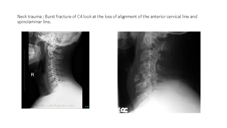
Neck trauma ; Burst fracture of C 4 look at the loss of alignment of the anterior cervical line and spinolaminar line.
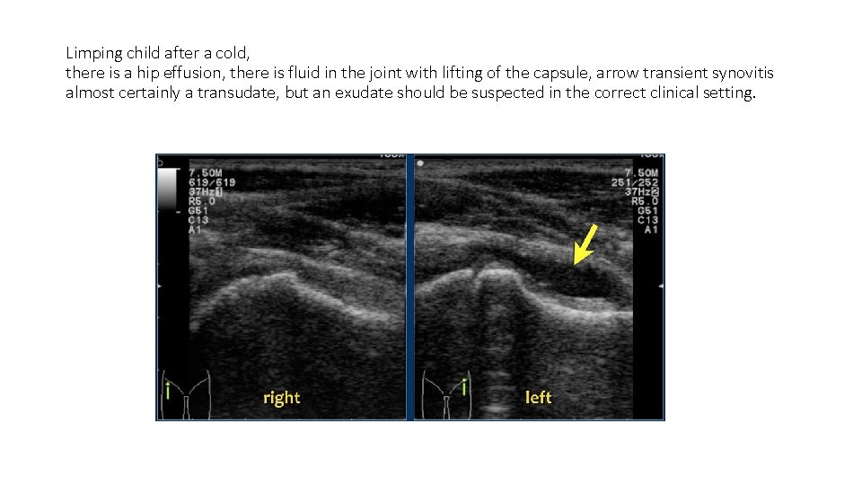
Limping child after a cold, there is a hip effusion, there is fluid in the joint with lifting of the capsule, arrow transient synovitis almost certainly a transudate, but an exudate should be suspected in the correct clinical setting.
THANK YOU