Paragonimus westermani Oriental lung fluke Paragonimus westermani causes
")


: ♠ Presented clinically by ♠")


")


 2 - Serological tests (IHA, IFA, ELISA): larvae trapped in human")


- Slides: 23
Paragonimus westermani (Oriental lung fluke)
Paragonimus westermani causes Paragonimiasis Geog. Distribution: Asia, Africa, South America. Encysted metacercaria Adult lives in cystic pockets of lungs Shrimps ﺟﻤﺒﺮﻯ Crabs ﺳﺮﻃﺎﻥ ﺍﻟﺒﺤﺮ Crayfish ﺟﺮﺍﺩﺍﻟﺒﺤﺮ
Paragonimus westermani 1 -Thick bodies . Reddish brown in color Oral sucker 2 - Adult fluke (10 x 5 mm) is ovate, rounded anteriorly caeca and slightly tapering posteriorly 3 - Cuticle is provided by spines 4 - Oral & ventral suckers are equal in size, the latter is situated near the middle of body 5 - Mouth leads to short esophagus & two simple intestinal ceacae. ovary testes 6 - Two irregularly lobed testes present side-byside in the posterior third of the body. uterus
Life Cycle of Paragonimus Infected man and fish-eating animal Mode of infection Ingestion of infected crabs, crayfish, shrimps Final host Cystic pockets in lungs Habitat Crab is the second intermediate host WATER Encysted metacercaria Infective stage Small tail Micro-cercous cercaria Egg in sputum or stool Diagnostic stage Semisulcospira Snail is the first intermediate host miracidium
• Life Cycle of Paragonimus
Development of Paragonimus inside Pathogenesis the body of infected human Granulomatous reactions around worms Fibrotic encapsulation Migration to ectopic sites adult Clinical Picture Chest pain. Chronic cough. Brownish sputum may contain eggs. Pleural effusion. Ectopic site Pleura Diaphragm Peritoneal cavity Ectopic site
Pathology & Clinical Picture ♠ Pathological findings depend on load of flukes & chronicity of infection ♠ Light infections are asymptomatic. ♠ In symptomatic infection, acute phase is cc by diarrhea, abdominal pain & urtecaria which correspond to invasion of intestine & subsequent migration of larvae to the lung ♠ These symptoms last for several wks ♠ Followed by fever, cough, night sweat, dyspnea & chest pain. ♠ Chronic phase infection may be pulmonary or extra pulmonary A- Pulmonary paragonimiasis Chronic Phase ♠ The most common form ♠ Lungs show congestion with reddish brown nodules ♠ Nodules composed of cysts containing adult worms usually in pairs & brownish, necrotic gelatinous exudates containing ova & Charcot Leyden crystals. ♠ Cyst wall is thick and contains granulation tissue with plasma cells, lymphoid cells, eosinophils & fibroblasts ♠ Bronchial mucosa is congested &oedematous.
Pathology & Clinical Picture A- Pulmonary paragonimiasis (cont. ): ♠ Presented clinically by ♠ Low grade fever. ♠ Cough with chocolate coloured sputum beaded with eggs ♠ Night sweat, severe chest pain, dyspnoea ♠ Recurrent attacks of bronchial pneumonia & haemoptysis. ♠ N. B. The condition mimics pulmonary TB B- Extra-Pulmonary paragonimiasis ♠ Migrating metacercariae may remain in peritoneal cavity or lodge in other organs e. g. liver, spleen, kidney, eye, brain, ….
Diagnosis I- Clinically: Chest pain, productive cough and diet history in endemic areas. II- Laboratory: 1 - Detection of eggs in sputum or faeces. 2 - Immunodiagnostic tests: detect early and chronic infection (ELISA and CFT ) 3 - Blood film examination to determine %age of eosinophila III- Plain X-ray of chest and computerized tomography.
1 - Detection of Paragonimus eggs In sputum or stool S : 90 X 55 µ S : oval, thick-shelled, opercutated at one pole, thickened at the other pole C : brown C : immature ovum 2 - Immunodiagnostic Tests a- Enzyme-linked Immunosorbent Assay (ELISA) Paragonimus Ag wash ELISA plate Patient’s serum Ab linked to enzyme wash substrate wash Coating well with Paragonimus Ag to detect anti. Paragonimus Ab in patient’s serum
Patient’s serum MAb wash Ab linked to enzyme wash substrate wash Coating well with anti-Paragonimus Ab to detect Paragonimus Ag in patient’s serum 2 - Immunodiagnostic Tests b- Complement fixation test (CFT) for antibody detection in patient’s serum Paragonimus antigen Complement Antibodies in patient’s serum Fixation of complement No Antibodies in patient’s serum No fixation of complement
Blood Film Examination Eosinophilia (20 -25%)
Plain X-ray Computerized tomography ﺗﺼﻮﻳﺮ ﻣﻘﻄﻌﻰ ﻳﻈﻬﺮ ﻋﻠﻰ ﺷﺎﺷﺔ ﺍﻟﻜﻤﺒﻴﺘﺮ Adult Paragonimus worms X-ray chest show patchy foci of fibrotic changes with ring shadows CT & MRI show cerebral cysts in cerebral paragonimiasis Treatment Praziquantel Adult worms may be expectorated after treatment
Epidemiology and Control Ø Epidemiology: Ingestion of raw or incompletely pickled, wine or brinesoaked infected crabs, crayfish & shrimps Pigs and small animals are paratenic hosts. Paratenic host: A host which carries the parasite in an arrested form. Ingestion of metacercariae during preparation of these fish via contaminated hands or utensils Ø Control: Treatment of cases. Proper cooking of crabs, crayfish and shrimps. Health education. Snail control.
Larva migrans Migration of larvae of nematodes in unsuitable hosts. Larvae cannot complete their normal development into adults. Cutaneous larva migrans Visceral larva migrans Invasion of human skin by filariform larvae of A. caninum & A. braziliense Invasion of human viscera by larvae of Toxocara canis & Toxocara cati
What happens when man ingests Larva is swallowed Embryonated A. lumbricoides egg ? 2 nd moult Rhabditiform larva Venous circulation Adult Ascaris in small intestine 3 rd & 4 th moults
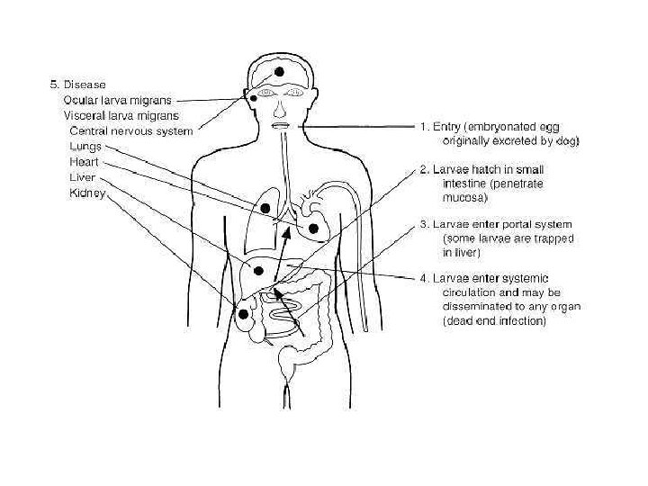
What happens when Man ingests Embryonated Toxocara canis egg? Toxocara cati egg? Visceral larva migrans Blood vessel Lymph vessel Eosinophilic granulomatous lesion
Pathogenesis and Clinical Picture of VLM n n Eosinophilic granulomatous lesions in viscera. lymphocytes Most frequently seen in the liver. eosinophils Foreign body giant cells Persistent eosinophilia with no symptoms. v Usual picture: Child – history of close contact with soil, dogs or cats. Larvae reaching the liver Enlarged tender liver. Larvae reaching the eye Visual disturbances. Larvae reaching the brain Neurological disturbances. Larvae reaching the lungs Pneumonitis. v
Diagnosis of Visceral Larva Migrans Clinical n Laboratory: 1 - Laparoscopy and biopsy of liver nodules. n
Diagnosis (cont. ) 2 - Serological tests (IHA, IFA, ELISA): larvae trapped in human tissues producing a state of chronic infection. This causes over-stimulation of the immune system producing increased amounts of: - Ig. M, Ig. G and Ig. E against larval Ag. - Anti-A and Anti-B isohaemagglutinins. Iso haemagglutinins: naturally occurring antibodies in patient’s serum against ABO group antigens on human RBCs. Larval antigens are similar to ABO antigens - Eosinophilia (20 - 80%)
Other causes of Visceral larva migrans In heavy infection: Occasionally, larvae of A. lumbricoides, A. duodenale or S. stercoralis fail to enter inside lung alveoli and are swept by the systemic circulation to settle in different organs of man. Larvae in arterial blood to viscera Larvae in venous blood
Treatment of Visceral Larva migrans 1 - Thiabendazole orally Repeated after one month 2 - Corticosteroids In severe cases Prevention and Control of Larva migrans Pet dogs and cats de-wormed. n Stray dogs and cats eliminated. Avoid contaminated food, drink and hands n