Nervous system Sensory organs Faculty of Pharmacy 12

- Slides: 40
Nervous system, Sensory organs Faculty of Pharmacy 12 th week histology practice Department of Anatomy, Histology and Embriology
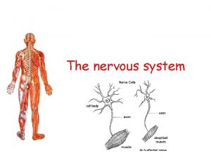
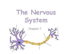
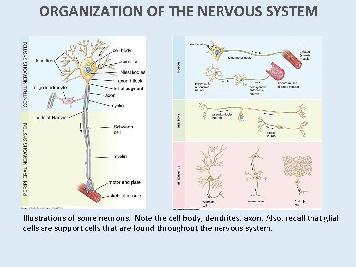
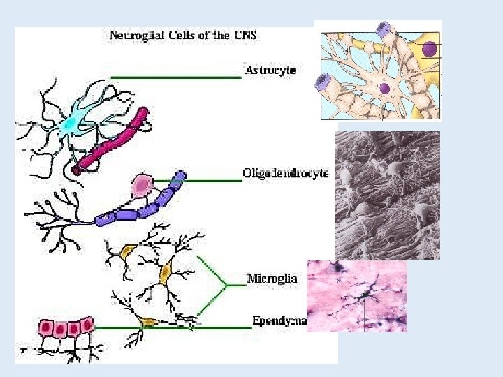
ORGANIZATION OF THE NERVOUS SYSTEM Illustrations of some neurons. Note the cell body, dendrites, axon. Also, recall that glial cells are support cells that are found throughout the nervous system.
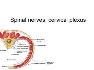
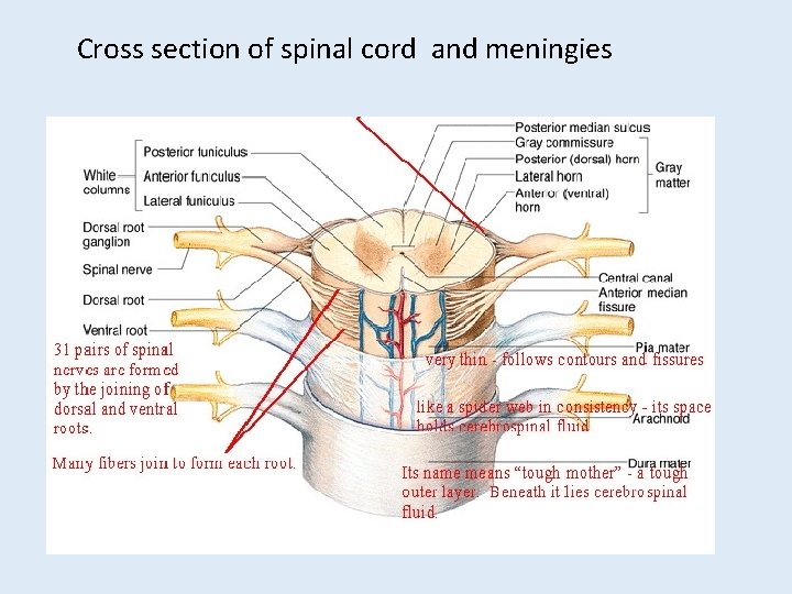
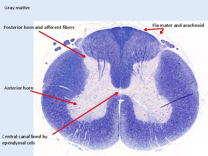
Cross section of spinal cord and meningies
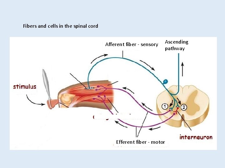
Fibers and cells in the spinal cord Afferent fiber - sensory Ascending pathway Efferent fiber - motor
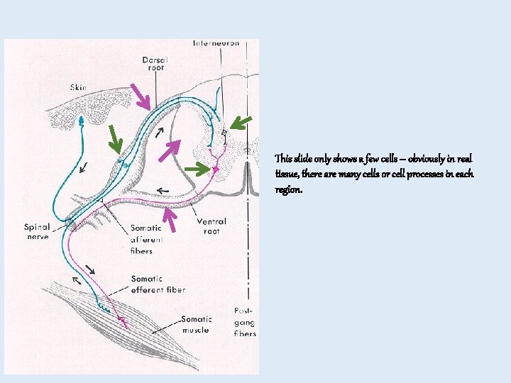
This slide only shows a few cells – obviously in real tissue, there are many cells or cell processes in each region.
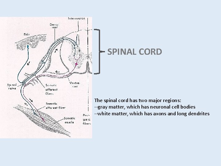
SPINAL CORD The spinal cord has two major regions: --gray matter, which has neuronal cell bodies --white matter, which has axons and long dendrites
Gray matter Posterior horn and afferent fibers Anterior horn Central canal lined by ependymal cels Pia mater and arachnoid
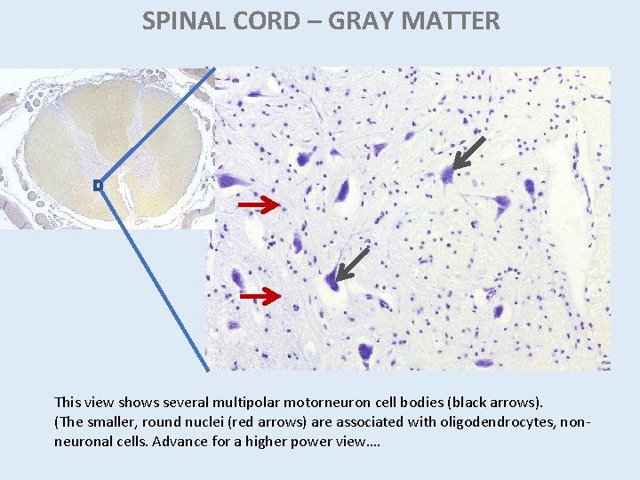
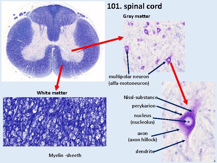
SPINAL CORD – GRAY MATTER This view shows several multipolar motorneuron cell bodies (black arrows). (The smaller, round nuclei (red arrows) are associated with oligodendrocytes, nonneuronal cells. Advance for a higher power view….
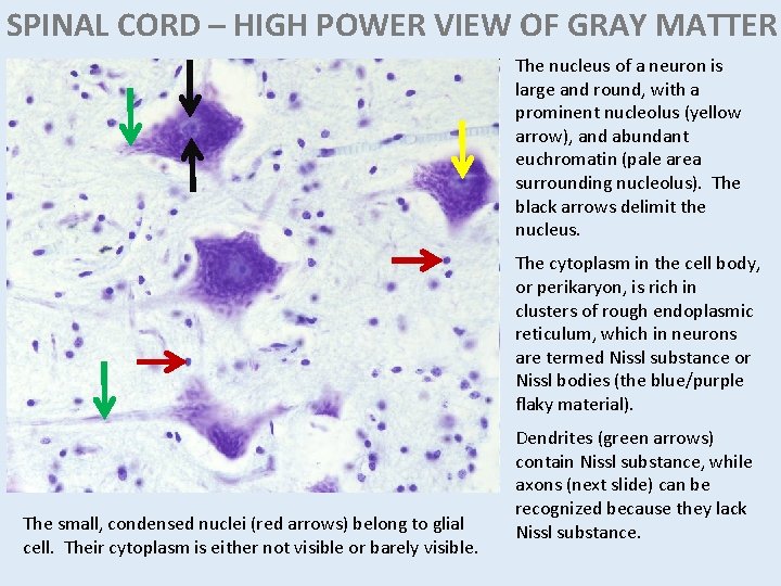
SPINAL CORD – HIGH POWER VIEW OF GRAY MATTER The nucleus of a neuron is large and round, with a prominent nucleolus (yellow arrow), and abundant euchromatin (pale area surrounding nucleolus). The black arrows delimit the nucleus. The cytoplasm in the cell body, or perikaryon, is rich in clusters of rough endoplasmic reticulum, which in neurons are termed Nissl substance or Nissl bodies (the blue/purple flaky material). The small, condensed nuclei (red arrows) belong to glial cell. Their cytoplasm is either not visible or barely visible. Dendrites (green arrows) contain Nissl substance, while axons (next slide) can be recognized because they lack Nissl substance.
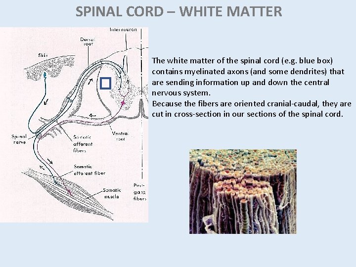
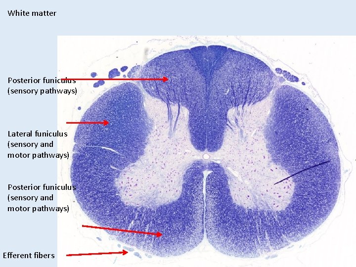
SPINAL CORD – WHITE MATTER The white matter of the spinal cord (e. g. blue box) contains myelinated axons (and some dendrites) that are sending information up and down the central nervous system. Because the fibers are oriented cranial-caudal, they are cut in cross-section in our sections of the spinal cord.
White matter Posterior funiculus (sensory pathways) Lateral funiculus (sensory and motor pathways) Posterior funiculus (sensory and motor pathways) Efferent fibers
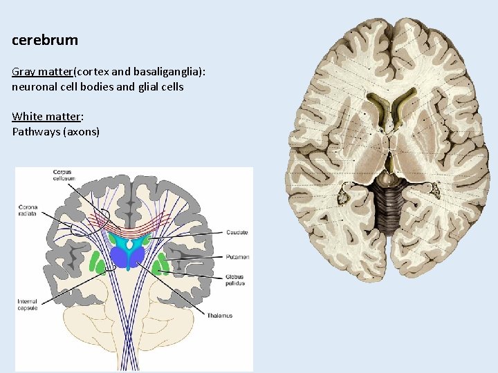
cerebrum Gray matter(cortex and basaliganglia): neuronal cell bodies and glial cells White matter: Pathways (axons)
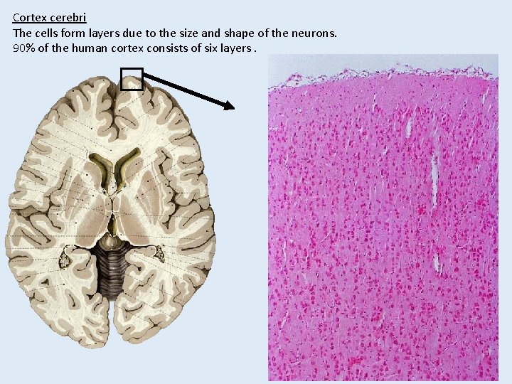
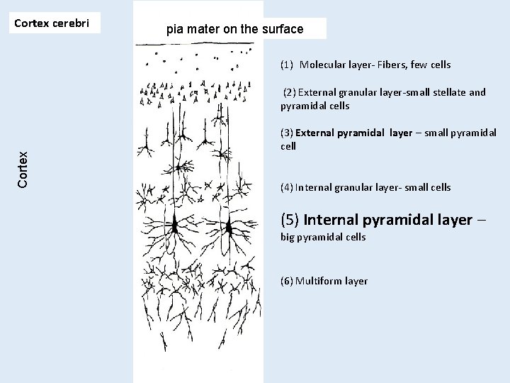
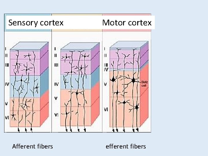
Cortex cerebri The cells form layers due to the size and shape of the neurons. 90% of the human cortex consists of six layers.
Cortex cerebri pia mater on the surface (1) Molecular layer- Fibers, few cells Cortex (2) External granular layer-small stellate and pyramidal cells (3) External pyramidal layer – small pyramidal cell (4) Internal granular layer- small cells (5) Internal pyramidal layer – big pyramidal cells (6) Multiform layer
Sensory cortex Afferent fibers Motor cortex efferent fibers
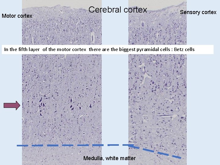
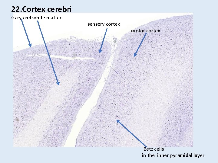
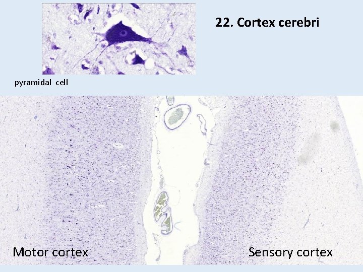
Motor cortex Cerebral cortex Sensory cortex In the fifth layer of the motor cortex there are the biggest pyramidal cells : Betz cells Medulla, white matter
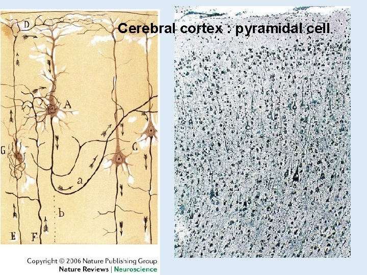
Cerebral cortex : pyramidal cell
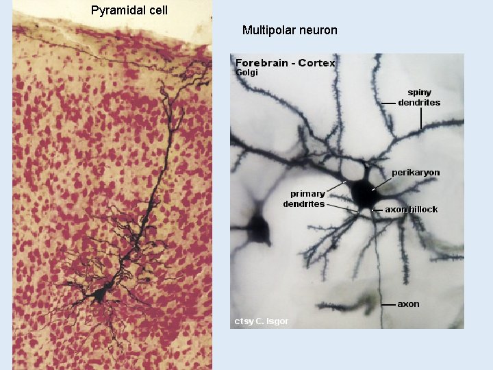
Pyramidal cell Multipolar neuron
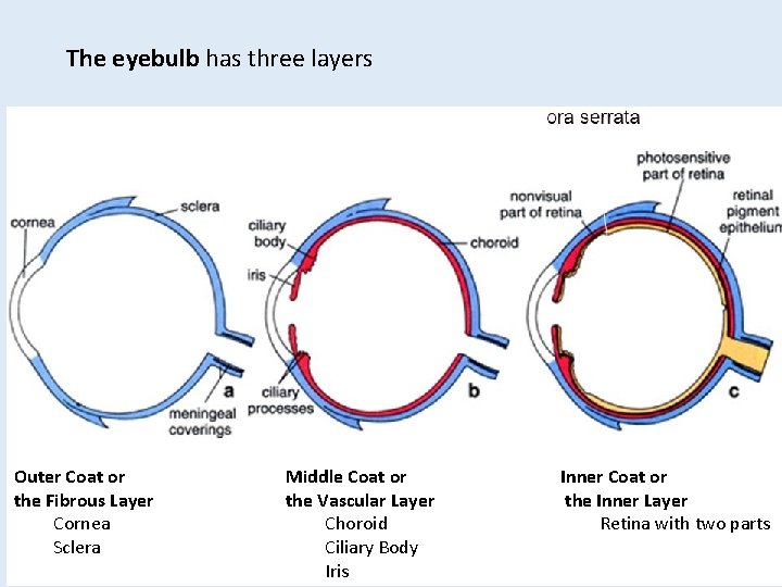
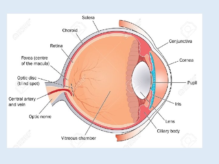
The eyebulb has three layers Outer Coat or the Fibrous Layer Cornea Sclera Middle Coat or the Vascular Layer Choroid Ciliary Body Iris Inner Coat or the Inner Layer Retina with two parts
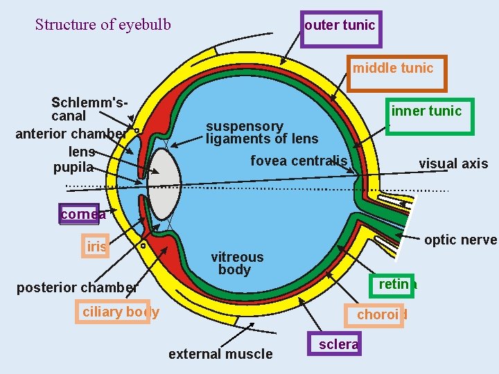
Structure of eyebulb outer tunic middle tunic Schlemm'scanal anterior chamber lens pupila inner tunic suspensory ligaments of lens fovea centralis visual axis cornea iris optic nerve vitreous body retina posterior chamber ciliary body choroid external musclera
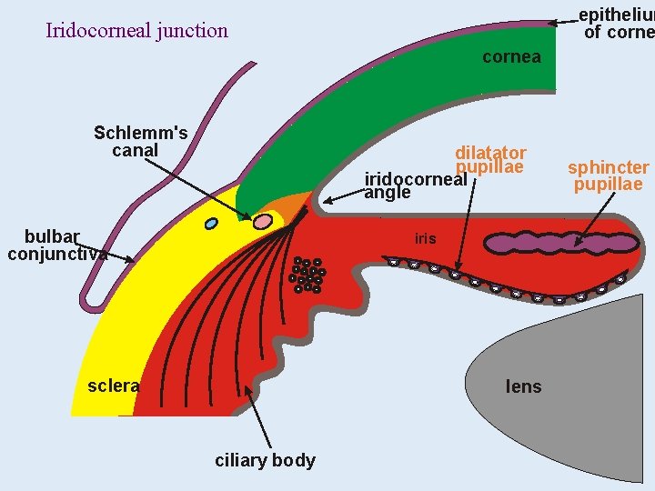
epithelium of corne Iridocorneal junction cornea Schlemm's canal dilatator pupillae iridocorneal angle bulbar conjunctiva iris sclera lens ciliary body sphincter pupillae
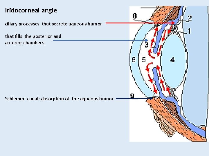
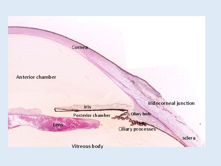
Iridocorneal angle ciliary processes that secrete aqueous humor that fills the posterior and anterior chambers. Schlemm- canal: absorption of the aqueous humor
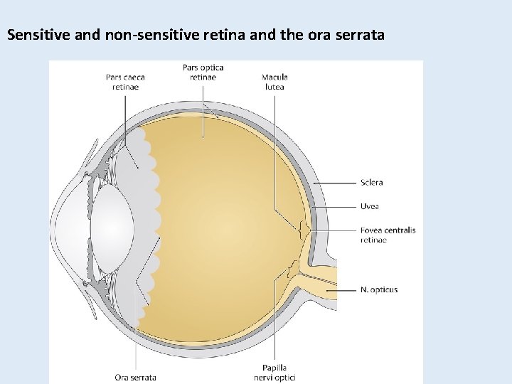
Sensitive and non-sensitive retina and the ora serrata
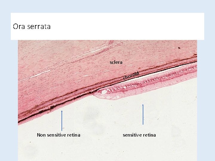
Ora serrata sclera d oi chor Non sensitive retina
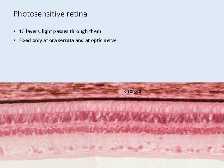
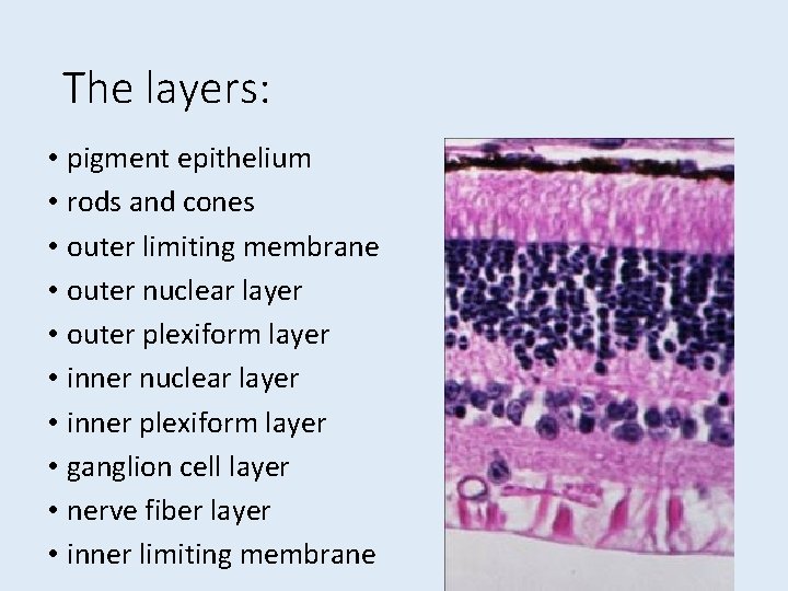
Photosensitive retina • 10 layers, light passes through them • Fixed only at ora serrata and at optic nerve choroid
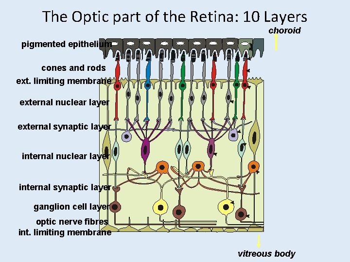
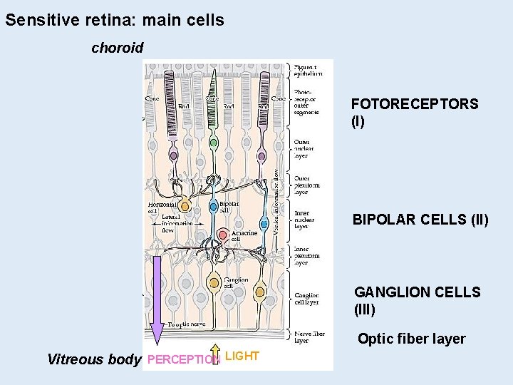
The Optic part of the Retina: 10 Layers choroid pigmented epithelium cones and rods ext. limiting membrane external nuclear layer external synaptic layer internal nuclear layer internal synaptic layer ganglion cell layer optic nerve fibres int. limiting membrane vitreous body
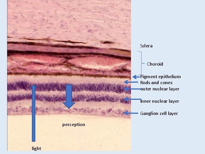
The layers: • pigment epithelium • rods and cones • outer limiting membrane • outer nuclear layer • outer plexiform layer • inner nuclear layer • inner plexiform layer • ganglion cell layer • nerve fiber layer • inner limiting membrane
Sensitive retina: main cells choroid FOTORECEPTORS (I) BIPOLAR CELLS (II) GANGLION CELLS (III) Optic fiber layer Vitreous body PERCEPTIONTLIGHT
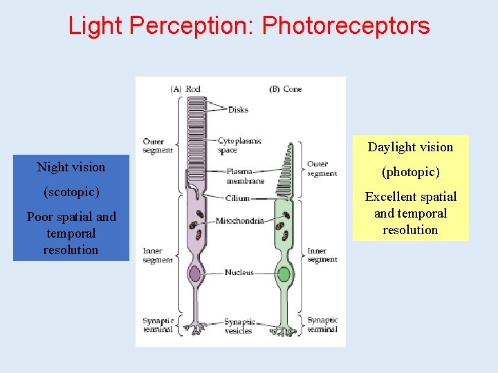
Light Perception: Photoreceptors Daylight vision Night vision (photopic) (scotopic) Excellent spatial and temporal resolution Poor spatial and temporal resolution
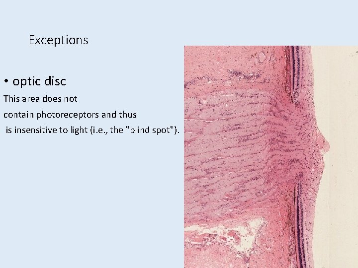
Exceptions • optic disc This area does not contain photoreceptors and thus is insensitive to light (i. e. , the "blind spot").
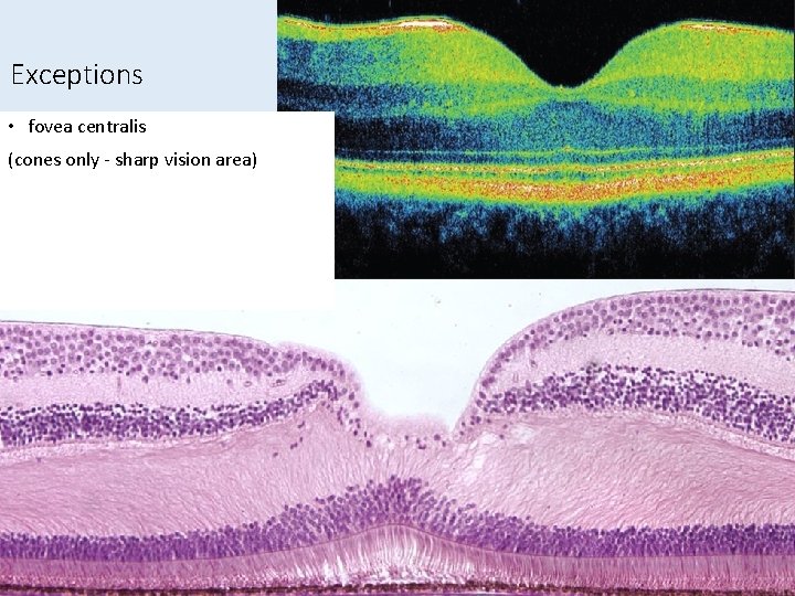
Exceptions • fovea centralis (cones only - sharp vision area)
Structures to identify
101. spinal cord Gray matter multipolar neuron (alfa-motoneuron) White matter Nissl-substance perykarion nucleus (nucleolus) axon (axon hillock) Myelin -sheeth dendrite
22. Cortex cerebri Gary and white matter sensory cortex motor cortex Betz cells in the inner pyramidal layer
22. Cortex cerebri pyramidal cell Motor cortex Sensory cortex
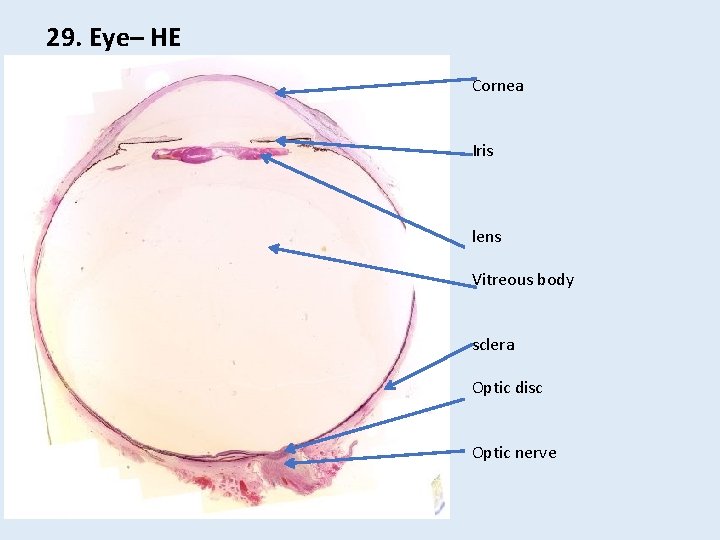
29. Eye– HE Cornea Iris lens Vitreous body sclera Optic disc Optic nerve
Cornea Anterior chamber iris Posterior chamber Lens Iridocorneal junction Ciliary body Ciliary processes sclera Vitreous body
Sclera Choroid Pigment epithelium Rods and cones outer nuclear layer Inner nuclear layer Ganglion cell layer perception light