Meiosis Topics 4 2 and 10 1 Assessment


 • Each")


- Slides: 25
Meiosis Topics 4. 2 and 10. 1
Assessment Statements 4. 2. 1 State that meiosis is a reduction division of a diploid nucleus to form haploid nuclei. 4. 2. 2 Define homologous chromosomes. 4. 2. 3 Outline the process of meiosis, including pairing of homologous chromosomes and crossing over, followed by two divisions, which results in four haploid cells. 4. 2. 4 Explain that non-disjunction can lead to changes in chromosome number, illustrated by reference to Down syndrome (trisomy 21). 4. 2. 5 State that, in karyotyping, chromosomes are arranged in pairs according to their size and structure. 4. 2. 6 State that karyotyping is performed using cells collected by chorionic villus sampling or amniocentesis, for pre-natal diagnosis of chromosome abnormalities. 4. 2. 7 Analyse a human karyotype to determine gender and whether non -disjunction has occurred.
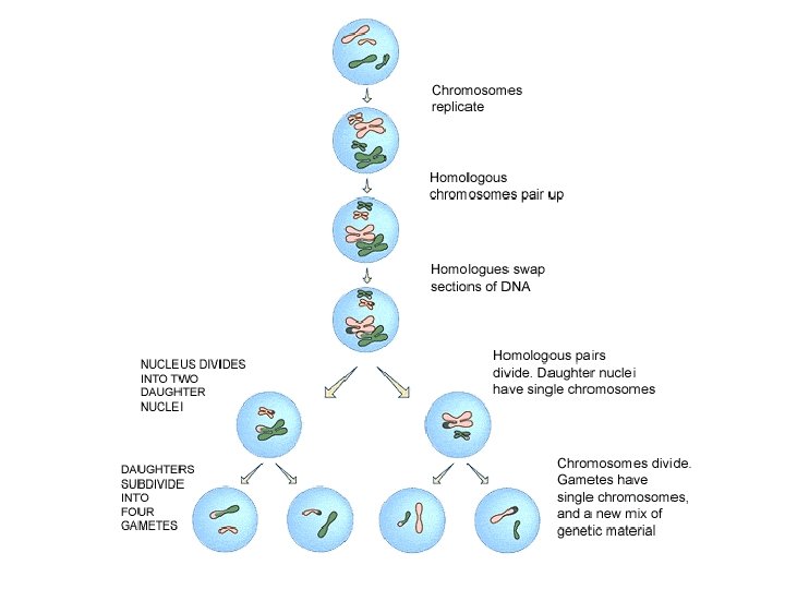
Meiosis • Form of cell division which results in gametes (sex cells) • Each new cell has only half the number of chromosomes that a typical cell in that organism has • These cells are haploid • Cells with the full number are called diploid cells • Division is called a reduction division because the number of chromosomes has been reduced • Four cells are formed and are called daughter cells • Each haploid daughter cell has a unique mix of half of the genetic information of the parent cell
Homologous chromosomes • Similar in shape and size • Two chromosomes carry the same genes • Not identical b/c the alleles could be different • The letter n is used to denote the number of unique chromosomes • Haploid (n) and diploid (2 n)
Phases of meiosis • Interphase • Prophase I – Chromosomes become visible – Homologous chromosomes pair up – Crossing over occurs – Spindle fibers form • Metaphase I – Homologues align across equator – Nuclear membrane disintegrates
• Anaphase I – Spindle fibers attach and pull chromosomes to poles • Telophase I – Spindles disintegrate – Chromosomes uncoil – New nuclear membranes form • Cytokinesis
• Prophase II – DNA condenses – New spindle produced • Metaphase II – Nuclear membrane disintegrates – Chromosomes align at equator – Spindle fibers attach to sister chromatids
• Anaphase II – Centromeres split, releasing each sister chromatid as an individual chromosome – Spindle fibers pull chromatids to poles • Telophase II – Chromosomes unwind – Nuclear envelopes from around each of the four haploid cells
Meiosis video Meiosis Square Dance
Down’s syndrome • Chromosomes do not always separate the way they are expected to do so during meiosis • Result is an unequal distribution of chromosomes due to non-disjunction • Non-disjunction of 21 st pair results in Down’s syndrome • A. k. a. Trisomy 21 • Effects: – Malformations of digestive system – Learning difficulties • Occurs in 1 in 800 births • Risk increases for mothers over 35
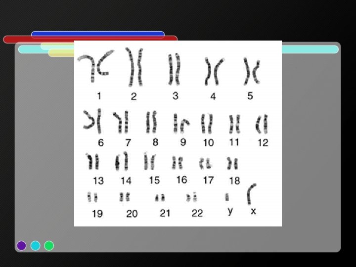
Karyotypes • 1. 2. 3. 4. Photograph of the chromosomes found in a cell Cells stained and prepared on slide Photomicrograph images obtained during mitotic metaphase Images cut out and separated Images place in order by size and the position of their centromeres
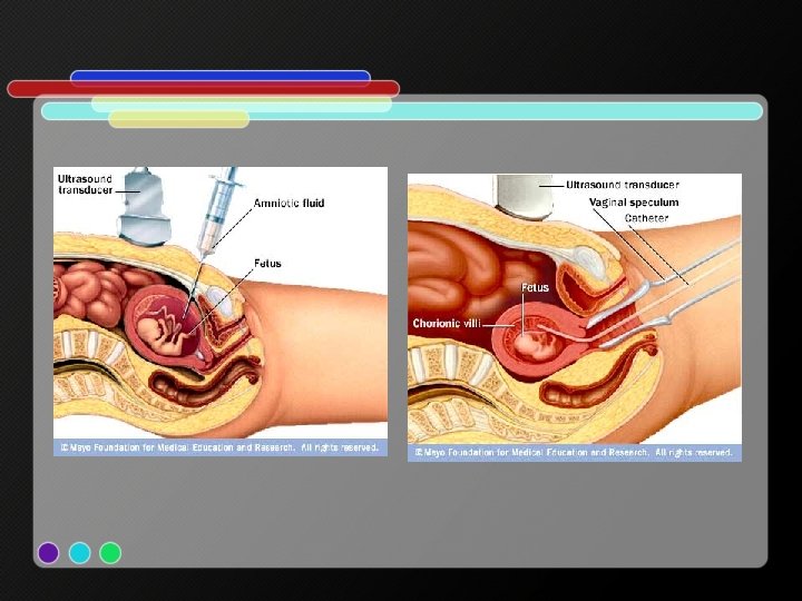
Obtaining cells for karyotyping • Amniocentesis – Hypodermic needle used to extract amniotic fluid around developing baby • Removing from chorionic villus – Obtained from tissue sample from the placenta’s projections into the uterine wall • White blood cells grown in lab • Very expensive and invasive • Recommended if expectant mother is over 35
Analysis of a human karyotype • • Gender? Non-disjunction? Karyotyping activity http: //www. biology. arizona. edu/human_bio /activities/karyotyping. html
Meiosis Topic 10. 1
Assessment Statements • 10. 1. 1 Describe the behaviour of the chromosomes in the phases of meiosis. • 10. 1. 2 Outline the formation of chiasmata in the process of crossing over. • 10. 1. 3 Explain how meiosis results in an effectively infinite genetic variety in gametes through crossing over in prophase I and random orientation in metaphase I. • 10. 1. 4 State Mendel’s law of independent assortment. • 10. 1. 5 Explain the relationship between Mendel’s law of independent assortment and meiosis.
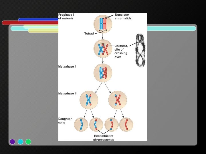
Chromosome behavior during meiosis Prophase I: chromosomes become more visible, homologous chromosomes pair up (called synapsis), crossing over occurs Prophase II: DNA condenses Metaphase I: centromeres have microtubules attached, bivalents line up randomly (random orientation), chromatids no longer identical Metaphase II: individual chromosomes line up along equator (random orientation), spindle fibers attach to centromeres Anaphase I: homologous chromosomes separate, pulled to opposite poles resulting in independent assortment Anaphase II: centromeres split, each sister chromatid becomes an individual chromosome and is pulled to opposite pole of cell Telophase I: chromosomes surrounded by two new nuclear membranes Telophase II: chromosomes unwind
Crossing over • Explains family resemblances • Adds chance for variety in offspring • Mixing genetic material between non-sister chromatids occurs when chromosomes intertwine and break • Tips of two chromatids are switched • The place where the two connect to each other is called a chiasma (pl. chiasmata)
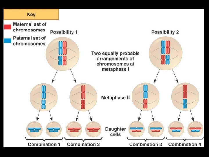
Genetic variety • Generated by: 1. Crossing over during prophase I • Chromatids which were once identical now have different combinations of maternal and paternal alleles at their various loci 2. Random orientation during metaphase I • Bivalents arrange themselves independent of chromosomes from same parent
Mendel’s law of independent assortment • When gametes are formed, the separation of one pair of alleles between the daughter cells is independent of the separation of another pair of alleles • Just b/c one trait (flower color) is inherited from a parent it does not follow that any specific trait of that parent (such as seed color) must be passed on as well • But, of course, there are exceptions to every rule… – This rule doesn’t always apply when genes are on the same chromosome
Independent assortment and meiosis • The distribution and separation of the chromosomes at the end of metaphase I ensures a random distribution of alleles between the gametes