Intraspinal Anomalies in Infantile Idiopathic Scoliosis Prevalence and


- Slides: 13

Intraspinal Anomalies in Infantile Idiopathic Scoliosis: Prevalence and Role of MRI Joshua M. Pahys, MD Amer F. Samdani, MD Randal R. Betz, MD Shriners Hospital for Children Philadelphia, PA, USA

Background • Reported prevalence of intraspinal anomalies in Infantile Idiopathic Scoliosis (IIS) is >20%

Background • Lewonowski et al. Spine 1992 – 2/4 (50%) IIS patients had Chiari malformations • Gupta et al. Spine 1998 – 3/6 (50%) IIS patients had intraspinal anomalies – 2/3 (66%) required surgical intervention

Background • Dobbs et al. JBJS 2002 – 10/46 IIS patients (21. 7%) had intraspinal anomalies – 8/10 (80%) required surgical intervention – Recommended screening MRI for all IIS patients with curves >20º

Purpose • To report the prevalence of intraspinal anomalies in patients with presumed Infantile Idiopathic Scoliosis routinely screened at a single, large volume institution. • Further define the role for a screening MRI in this patient population.

Intraspinal Anomalies Syrinx Tethered Cord Chiari

Intraspinal Anomalies • 7 of 54 patients (13%): Positive MRI • Tethered Cord: 3 patients • Chiari Malformation: 2 patients • Nonoperative Syrinx: 2 patients • 5 of the 7 patients (71. 4%): required neurosurgical intervention

Results Sex Age at Presentation (months) 27 Female (57. 4%) 14. 3 20 Male (42. 6%) (range: 4 -34) 4 Female (57%) 10. 7 3 Male (43%) (range: 1 -30) 0. 98 0. 21 Normal MRI Abnormal MRI p-value (Normal vs. Abnormal)

Results Main Curve Apex Location Main Curve Direction Main Curve Cobb Angle 41 Thoracic (87. 2%) 37 Left (78. 7%) 49. 1° 6 Lumbar (12. 8%) 10 Right (11. 3%) range: 20°-106° Abnormal MRI 6 Thoracic (85. 7%) 4 Left (57. 1%) 48. 4º 1 Lumbar (14. 3%) 3 Right (42. 9%) range: 22°-90° 0. 92 0. 22 0. 94 Normal MRI p-value (Normal vs. Abnormal)

Results Normal MRI Abnormal MRI Curve Magnitude (patients) 20º-29º 30º-39º 40º-49º > 50º 20 9 14 4 ( 19. 1%) (29. 8%) (8. 5%) 2 1 (28. 6%) (14. 3%) 1 (14. 3%) (42. 6%) 3 (42. 9%)

MRI in Infants • Sedation/General Anesthesia often required • Malviya et al. Anesth Analg 1997 – Sedation for 1, 140 infant MRI’s – 20% incidence of adverse events – 5. 5% incidence of hypoxemia • Malviya et al. Br J Anaesth 2000 – Sedation for 922 infant MRI’s – 7% failed scans secondary to inadequate sedation

When to get an MRI? • “Curve progression should be the major indication for a magnetic resonance imaging scan in patients with early onset scoliosis. ” – Fernandes/Weinstein JBJS 2007 • Our recommendations: – Curve progression >10º per year – Change in neurologic exam – Surgical intervention planned

Conclusion • A smaller percentage (13%) of neural axis abnormalities was identified in this population than previously reported • A screening MRI may not be necessary in all patients at presentation with infantile idiopathic scoliosis measuring >20°
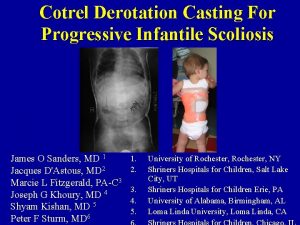
 Infantile scoliosis casting
 Period prevalence vs point prevalence
 Period prevalence formula
 Period prevalence vs point prevalence
 Period prevalence vs point prevalence
 Ideopathic peripheral neuropathy
 Idiopathic hirsutism
 Idiopathic oat
 Pediatric surgery
 Rvad spine
 Scoliosis chiropractor seminole county
 Postural screening worksheet
 Scoliosis
 Scoliosis research society


