Immunofluorescence in Dermatopathology DR AZADEH RAKHHAN DERMATOPATHOLOGIST SHOHADAETAJRISH

 is a histochemical technique employed to detect antibodies bound to antigens")

: DIF is a one step procedure that involves application of")


 Type of immunoreactant: Ig. G, Ig. A, Ig. M,")


 and")
 deposits at")


 is subepidermal bullous disease of unknown cause. Major antigens in BP")


- Slides: 63
Immunofluorescence in Dermatopathology DR. AZADEH RAKHHAN DERMATOPATHOLOGIST SHOHADA-E-TAJRISH HOSPITAL
Immunofluorescence (IF) is a histochemical technique employed to detect antibodies bound to antigens in the tissue or in the circulating body fluids. It acts as a valuable adjunct to clinical and histopathological diagnosis, especially in vesiculobullous and connective tissue disorders. Coons in 1940 s was first to apply the IF technique to demonstrate the microorganism in the infected tissue. Its application in dermatopathology came much later; in 1963, when these techniques were used to demonstrate the deposition of Ig and complement at the DEJ in SLE (”lupus band test”)
Principle IF technique involves viewing of antigen–antibody complexes under ultraviolet microscope using corresponding antibodies tagged to a fluorochrome. Fluorochromes are compounds containing electrons which when irradiated with a light of a particular wavelength achieve an unstable higher energetic state. On returning to the ground state as a spontaneous process, they emit light of a longer wavelength. Simple conjugation process, retention of the antibody activity in the labeled protein, and stability of the fluorescent conjugate are prerequisites of an ideal fluorochrome. Fluorochromes, currently in use, are fluorescein isothiocyanate (FITC) which produces apple-green color and tetramethylrhodamine isothiocyanate (TRITC) with a red color of fluorescence
Direct immunofluorescence (DIF): DIF is a one step procedure that involves application of fluoresceinated antibodies to a frozen section of the skin. This test determines the deposition of immunoreactants in the patient's tissue. Indirect immunofluorescence (IIF): In IIF, normal whole tissue is the substrate (usually monkey esophagus). This method requires two incubations. The patient's serum is layered on the substrate followed by application of fluoresceinated antibodies. These studies detect circulating antibodies in the serum. An important advantage of the indirect method is its increased sensitivity (10 -15 times). A modified IIF technique using the patient's own skin as a substrate known as immunomapping (antigen mapping) is used to determine the exact site of cleavage or abnormalities in the distribution of mutated structural proteins (normal, reduced, or lack of expression) in various forms of hereditary epidermolysis bullosa (EB). Complement fixation: This is another type of IIF. After the patient's serum is layered on the substrate, a source of complement is added. Fluoresceinated anticomplement antibodies are then used to detect the presence of complement in the tissue. This test can detect small quantities of complement fixing antibodies. Immunoelectron microscopy (IEM): It can be performed in an analogous fashion to detect DIF or IIF. Instead of fluoresceinated antibodies, the antibodies are labeled with an enzyme, such as horseradish peroxidase or a heavy metal, such as colloidal gold. This test provides subcellular or ultrastructural localization of immunoreactants. This technique may be helpful in the differential diagnosis of subtypes of hereditary EB, where antigen mapping is not significant. There are several advantages of IF over immunoelectron microscopy. IF is a technically simpler and shorter procedure than IEM, and it is more quantitative and reliable. It is also less costly in terms of technician time and reagents; however, tests are less permanent than those made with peroxidase staining. Another major difference is that the biopsy requires specialized cutting of thin sections for immunoelectron microscopy.
Types of specimens Skin biopsy specimen: for in vivo deposits of serum proteins, usually by the DIF staining method Serum samples: About 3 ml of blood without anticoagulants are collected, and the serum is separated from the clotted blood. Clotted blood samples should not be frozen as this causes massive hemolysis. Serum samples are tested for circulating autoantibodies by IIF. For diagnosis of hereditary EB, about 2 -5 ml of ethylenediaminetetraacetic acid (EDTA) blood is needed for mutation analysis. For any "IF" method, direct or indirect, basically, a section of tissue sample or cellular smear fixed on a glass slide, known as "substrate", is used. In addition, a "conjugate", i. e. an antiserum (e. g. against immunoglobulins, complement components, and fibrinogen) that is covalently linked to a fluorochrome, is required.
Site of skin biopsy Skin biopsy is taken from the unblistered perilesional (normal looking skin) area of the fresh lesion since in older lesions the basement membrane may be completely destroyed, resulting in a negative IF test. It is advantageous to include parts of a fresh blister to get information about the level of split formation. If the biopsy is taken only from the lesional skin, IF again will be negative as in vivo-bound autoantibodies may have been removed by the inflammation. For lesions on mucous membranes, biopsy for immunologic studies should be taken from the periphery of a fresh lesion, since the epithelium in old lesions is usually extensively eroded and therefore the immunofluorescent reaction cannot be detected.
Dermatitis herpetiformis : The standard practice is to obtain biopsy specimens from normal-appearing perilesional skin as inflammation in lesional skin degrades the immune reactants and is usually falsely negative for the diagnostic granular pattern. Extreme degree of precaution is highly recommended while taking a punch biopsy for hereditary EB as the skin in these patients is extremely fragile and the biopsy procedure itself leads to dermo-epidermal separation causing retention of epidermis within the punch instrument during the procedure.
Ø Lupus erythematosus Biopsies should be taken from both lesional and apparently normal skin in sun-exposed areas. As a general rule, sun-exposed lesional skin should be used to substantiate an initial diagnosis of LE so as to avoid the problem of false-negative results due to decreased sensitivity in sun-protected areas. In discoid lupus erythematosus (DLE), biopsy should be taken exclusively from lesional skin as uninvolved skin is usually negative.
Vasculitis : A freshly erupted purpuric spot in the most proximal part of the limb is preferred as Ig. A deposits may undergo degradation in older lesions. In polyarteritis nodosa, deep skin biopsies are needed as IF staining is evident only in some deep arteries in subcutis. Porphyria : Biopsies should be taken from the dorsum of hand lesions as well as normal skin. Lichen planus : Biopsy is taken from inflamed mucosa and/or skin.
Transportation of the Biopsy Sample Skin biopsy sample should be transported to the laboratory in phosphate-buffered saline (PBS). Normal saline is also shown as a useful transport medium if the samples can be shipped to the IF laboratory within 24 h. If the facility for IF is not available locally, biopsy sample can be transported to the test center in Michel's medium (MM). This transport medium contains ammonium sulfate, N-ethylmaleimide, potassium citrate buffer, magnesium sulfate, and distilled water. It probably preserves immunoantigenicity of the specimen by its ability to precipitate macromolecules while inhibiting proteolytic enzymes. Immunoreactants may be demonstrable by DIF even at 6 months, indicating the reliability of this medium in longterm preservations of skin biopsies.
DIF staining protocol A 3– 4 mm punch biopsy is optimum for DIF study; to get a maximum yield, it is important to take biopsy from an appropriate site. Lesional biopsy is also preferred in cases of : discoid lupus erythematosus (DLE) Amyloidosis lichen planus (LP) porphyria cutanea tarda (PCT) It is important to avoid contamination of biopsy samples with formalin which render the skin specimen unsuitable for DIF study. the first biopsy should always be taken for DIF.
Cryocut sections must be dry before staining or they will detach and wash away. For staining, sections are brought to room temperature and washed in PBS to remove unbound serum proteins which can significantly contribute to background staining. Optimally diluted FITC-labeled monospecific immunoglobulins (Ig. G, Ig. A, and Ig. M), C 3 , C 5 b-9 and fibrin are layered onto the section while it is still moist and incubated at 37 °C for 45 min to 1 h. Then, the sections are washed in PBS thrice and mounted in buffered glycerin. Buffered glycerin at neutral p. H is the simplest and most commonly used mountant for IF slides. Rapid fading of fluorescence after excitation is one of the most serious disadvantages of IF techniques, which limits the re-examination of slides for further interpretation. The preservation of fluorescence in DIF-positive slides using mounting media with an antifading reagent is possible for 2 years at room temperature; however, in daily practice, storage for longer than 11 months prevents a reliable diagnosis. Therefore, photography should always be used to document the results.
Skin biopsy cannot be kept beyond 1 month in Michel's fluid at 4 -8 °C. Once frozen sections of skin biopsies are cut and layered onto the slides, they can be stored indefinitely in deep freezers. During this storage period, thawing of the sections should be absolutely avoided as thawing makes the slides unfit for DIF reporting ( uninterrupted electrical supply is needed) Two sections are layered on each slide. The slides are then air dried and stored at − 20 °C until they are stained. Before starting the staining procedure, the sections have to be thawed and air dried for at least 2 h at room temperature to prevent floating away of the sections while staining. Freshly stained biopsy should be stored away from light at 4 -8 °C and be reported on the same day to prevent quenching of fluorescence. Ideally, digital recording of findings should be maintained.
Sections of 4– 6 µm thickness cut using a cryostat adhesive slides Sections are then air dried and washed in PBS to remove any unbound proteins. It is then treated with adequately diluted FITC-labeled conjugates (Ig. G, Ig. M, Ig. A, C 3, and fibrin) and incubated for 1 h in a moist chamber at room temperature. Alternatively, slides can be coated with poly-L-lysine to improve the adhesive property. The sections are then washed in PBS (three washes of 10 min each) and mounted in buffered glycerol and examined under fluorescent microscope.
Serum samples All sera should be refrigerated until tests are performed. During frozen storage for many months or years, antibody levels decline gradually. Repeated freezing and thawing should be avoided, since this causes a rapid loss of antibody activity. In general, one or two freeze-thaw cycles can be tolerated, but five or more can cause almost complete loss of activity. Positive and negative control sera must be frozen in aliquots of a size adequate for single experiment. EDTA blood should not be frozen and if this cannot be assured, it is recommended to extract DNA and then store it at -80 °C.
IIF staining protocol The substrate is incubated with a series of dilutions of patient serum in PBS (mostly starting with 1: 10) for 30 min at room temperature and then washed. If present, circulating autoantibodies will bind to the respective antigens in the substrates and are detected by incubation with FITC-labeled, mouse antihuman Ig. G and / or Ig. A. Screening for ANA should be done at dilutions of 1/40, because lower dilutions give too many false positives. Even a dilution of 1/40 gives about 20% of ANA positives with normal sera.
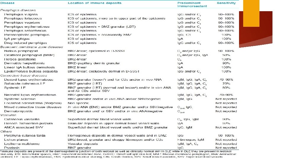
Reporting the Skin biopsies 1) Type of immunoreactant: Ig. G, Ig. A, Ig. M, C 3, fibrin, C 5 b-9. 2) Location of immune deposits: intercellular spaces (ICS) in epidermis/epidermal nuclear staining (ENS) or in vivo ANA/basement membrane zone (BMZ)/subepidermal blood vessels/hair shaft/cytoid (civatte or colloid) bodies. 3) Extent of staining: Focal or diffuse. 4) Intensity of staining: Semi-quantitative grading of strength of fluorescence: + to ++++. 5) Pattern of immune complex deposits: granular or linear. The granular pattern is further divided into coarse granules, speckles, threads, and fibrils.
Key points to remember while reporting There is often a need to compare relative intensities of staining between positive and background staining in control slides. immunopathologist must also be aware of nonspecific staining and autofluorescence. High background often results from antibodies that are too heavily conjugated with fluorescein; these molecules adhere nonspecifically to tissue because of their negative charge. It may also be the result of partial drying of tissue sections after the staining procedure has begun. Nonspecific staining is fluorochroming due to staining capacity of FITC-labeled proteins. It depends on the affinity of the specific components of the antigenic substrate for nonspecific binding of labeled protein and the concentration of fluorescein in the dilution of conjugate selected for staining. Nonspecific staining is commonly encountered in thicker sections. Sections cut at 6 μm or thicker cannot be used for IF because of high nonspecific staining. Autofluorescence, due to fluorescence found intrinsically in the tissue (recognized usually by its color which is generally yellow or orange), should be distinguished from the apple green color of fluorescein. Examination of an unstained (control) section can be helpful in judging autofluorescence.
Slides should be examined preferably on the day of staining and a detailed record of results as outlined above should be kept on work sheets. It is wise to run positive and negative controls with each biopsy, until one gains confidence in specificity of staining with a given conjugated antibody.
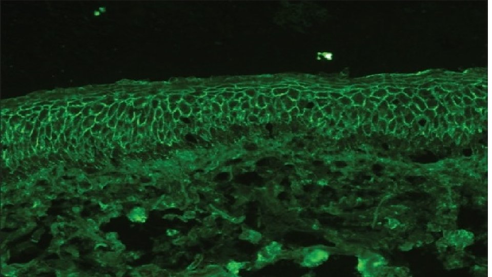
1 - Ig. G staining in Intercellular space Autoantibodies in pemphigus are directed against desmosomal proteins, namely desmoglein 1 and 3 (Dsg 1 and 3) which are responsible for cell-to-cell adhesion in the epidermis. This pattern is seen in all types of pemphigus except Ig. A pemphigus. The staining pattern is identical in pemphigus vulgaris (PV) and pemphigus foliaceus (PF), but at times, the fluorescence may be localized to or more intense along the upper layers of epidermis in PF. C 3 deposition follows the same pattern as Ig. G, but it is less intensely stained compared to Ig. G and usually detected in patients with active disease. “chicken-wire” or “fish-net” appearance. The sensitivity of DIF is around 90%– 100% in patients with active disease.
pemphigus DIF of perilesional skin in pemphigus shows linear/granular deposition of immunoglobulins (Igs) and C 3 in the epidermal ICS: "chicken wire" pattern. All forms of pemphigus produce the same pattern, though there is some tendency for staining to be more superficial within the epidermis in PF than in PV. In pemphigus erythematosus, a mixed pattern is seen, i. e. ICS positivity with a band of immune deposits along the dermo-epidermal junction (DEJ). Ig. G is present in nearly 100% of patients whereas C 3 is present in 50 -100% of cases. The intensity of DIF coincides fairly well with clinical activity, but a few patients may continue to have positive DIF despite clinical inactivity of pemphigus. In patients who relapse, DIF tends to remain positive and the complement deposition may show an increase or may reappear. It is, thus, a better indicator of an imminent relapse. The pemphigus-specific immunofluorescence pattern seen in the skin has also been demonstrated in the outer root sheaths of plucked hair follicles in 85 -100% cases of PV. Tzanck smears can serve as an alternative to surgical biopsies especially in rural areas lacking proper histological equipment. IIF studies on appropriate epithelial substrates detect circulating Ig. G autoantibodies, which produce the characteristic ICS pattern of fluorescence. The presence of these autoantibodies is abnormal at any titre and generally correlate with disease activity. ELISA (desmoglein 1 and 3) is a quick, simple, sensitive, and efficient tool for confirming the diagnosis as well as predicting relapse
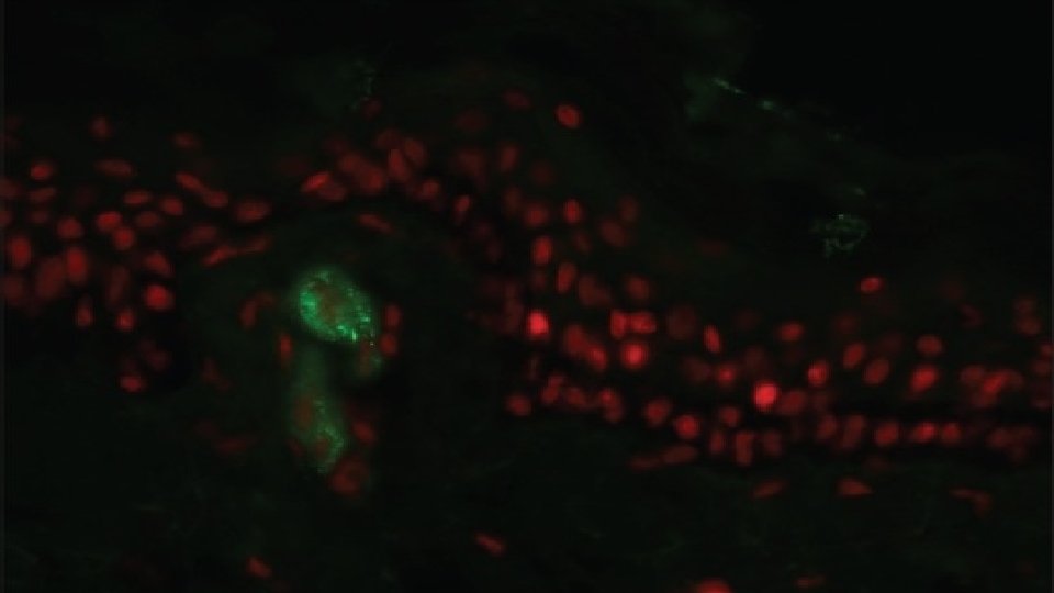
DIF of a case of PV showing Ig. G reactive granular (+++) deposits at the intercellular spaces in the epidermis
2 -Ig. A staining in intercellular space Ig. A pemphigus is characterized by Ig. A deposition in the ICS two types of Ig. A pemphigus have been recognized - subcorneal pustular dermatoses (SPD) type and intraepidermal neutrophilic (IEN) type. In SPD type, Ig. A deposition is seen predominantly in the upper epidermal layers in IEN type, it is seen throughout the epidermis or restricted to the lower epidermis.
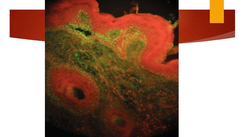
3 -Intercellular and basement membrane zone staining This type of dual staining of epidermal ICS and basement membrane zone (BMZ) occurs in two conditions: pemphigus erythematosus (PE) paraneoplastic pemphigus (PNP) PE is a variant of PF characterized by immunopathological coexistence of PF and lupus erythematosus (LE). DIF in PE reveals ICS in a “fish-net” pattern; in addition, there is granular BMZ staining with Ig. G resembling “lupus band. ” Occasionally, these patients may have circulating antinuclear antibodies (ANA) in their blood.
PNP is characterized by autoantibodies against desmosomal (Dsg 1 and 3, desmoplakin, envoplakin, and periplakin) as well as BMZ protein (bullous pemphigoid [BP]. DIF in PNP reveals ICS and linear BMZ staining with Ig. G and C 3. Intercellular staining in PNP tends to be weak, diffuse, and nonspecific, while deposition along BMZ is almost identical to that of BP
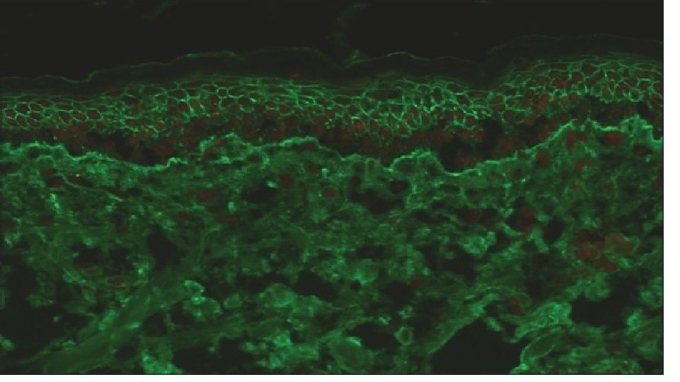
4 -Linear basement membrane zone staining-Ig. G The deposition of Ig. G, C 3, or both in a linear fashion along the BMZ is seen in BP, mucous membrane pemphigoid (MMP), PG, epidermolysis bullosa aquistia (EBA), bullous SLE, and recently described anti-p 200 pemphigoid. Relative intensity of staining with Ig. G and C 3 may sometimes help to provisionally subcategorize these conditions. For example, a more intense staining of BMZ with C 3 when compared to Ig. G indicates the diagnosis of pemphigoid group of disorders (BP, MMP, and PG); vice versa holds good for EBA. PG is characterized by an exclusive staining of BMZ with C 3. presence of multiple deposits at BMZ strongly suggests the diagnosis of EBA or bullous SLE. In EBA, intense Ig. G deposition is almost always present. The intensity of C 3 deposits is less than Ig. G. Ig. M is found in half of the cases, and Ig. A is found in twothird cases of EBA. In the absence of clinical history, it may be impossible to differentiate between EBA from bullous SLE as DIF features may be similar in these conditions.
Bullous pemphigoid (BP) is subepidermal bullous disease of unknown cause. Major antigens in BP are two keratinocyte hemidesmosomal proteins, an intracellular 230 k. Da protein (BP 230) and 180 k. Da protein (BP 180). DIF of unblistered, perilesional skin from the edge of a fresh blister is diagnostically sensitive and typically demonstrates linear BMZ fluorescence in nearly 100% of patients. C 3 and Ig. G, or C 3 alone are the predominant immunoreactants. A linear BMZ pattern can be seen in several disparate clinical disorders, including epidermolysis bullosa acquisita (EBA) and gestational pemphigoid. In BP and gestational pemphigoid, linear Ig. G is localized to the roof (epidermal side) of the split whereas in EBA, linear deposition of immunoreactants is seen along the dermal side of the split. IIF detects circulating Ig. G anti-BMZ autoantibodies in about 50% of the patients with pemphigoid, but the sensitivity increases to 80% if normal human salt-split skin is used as a substrate. The combined ELISAs based on BP 180 and BP 230 fragments are very useful in establishing the diagnosis of BP and detect Ig. G autoantibodies in 96% of all BP sera and thus can be used as adjunctive diagnostic aid
C 3 in BP
Gestational pemphigoid It develops during or soon after pregnancy. DIF from perilesional skin typically shows linear C 3 BMZ deposits in 100% of cases. Ig. G may be seen in 30% cases.
Epidermolysis bullosa acquisita It is an acquired subepidermal blistering disease characterized by the presence of autoantibodies to type VII collagen in BMZ. DIF of direct salt-split skin biopsy tissue shows linear deposits of immunoreactants at the dermal, or floor aspect of the induced blister. On IIFM, only about 40% of EBA patients have demonstrable antibodies.
5 -Linear basement membrane zone staining-Ig. A Linear Ig. A bullous dermatosis In nearly 100% cases, DIF demonstrates intense linear Ig. A deposits along the BMZ, but none in dermal papillae. Weaker staining for Ig. G, Ig. M, and/or C 3 is also found in about 20% of cases. This pattern of staining is not entirely specific for linear Ig. A dermatosis as some cases of EBA that are caused by Ig. A anti-collagen VII antibodies produce a similar pattern on DIF. Linear Ig. A at the BMZ also occurs in 20% of mucous membrane pemphigoid cases. In these diseases, the Ig. A is often accompanied by other immunoreactants at the BMZ, but this can also occur in linear Ig. A disease, and there is some overlap among the three conditions. Linear or granular Ig. A can also be observed at the BMZ in lupus. In SSST, linear staining can be seen on roof or floor. On IIF, circulating Ig. A anti-BMZ autoantibodies can be detected in about 10 -30% cases.
An exclusive linear deposition Ig. A along the BMZ is a pathognomonic feature of linear Ig. A disease (LAD).
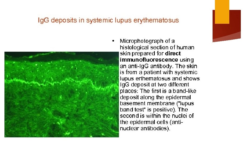
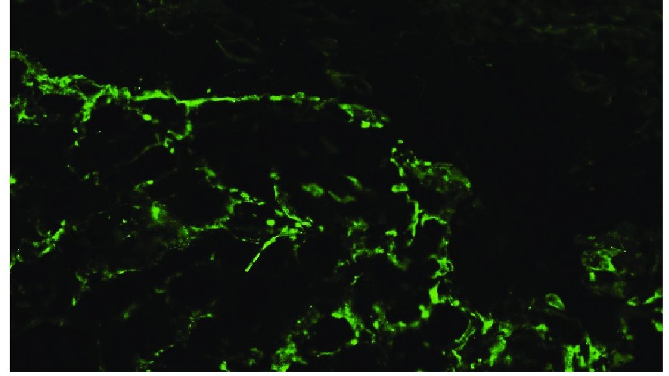
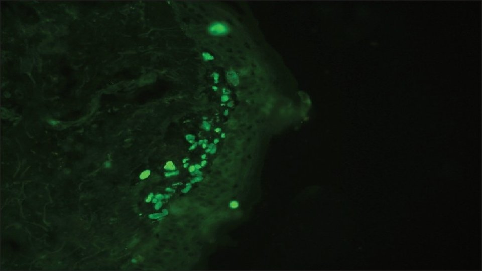
6 -Granular basement membrane zone staining SLE demonstrates the deposition of immunoreactants in a granular pattern along the BMZ both in the lesional skin and sun protected, nonlesional skin (”the lupus band test”). It is usually seen with Ig. M but may be seen with other immunoreactants as well. In fact, presence of 3 or more immunoreactants deposition in BMZ is highly suggestive of SLE. In addition, colloid bodies (staining frequently with Ig. M) in the papillary dermis and epidermal nuclear staining with Ig. G (epidermal “ANA”) may be seen in SLE. DLE, reveals BMZ staining with these immunoreactants which may be more homogeneous and thick, especially when the biopsy is taken from the well-established lesion.
Lupus erythematosus characteristic coarse, granular, continuous deposits of Igs and complement along the DEJ (LBT). The reported sensitivity of DIF examination for lupus ranges from 58% to 93%. Although characteristic, this reaction pattern can be seen in other disorders such as mixed connective tissue disease, dermatomyositis, scleroderma, drug eruptions, and vasculitis. Therefore, it is mandatory to correlate DIF results with both clinical presentation and histopathological findings. Up to 95% of individuals with DLE show LBT. However, positive DIF findings in DLE are confined to lesional skin, whereas skin with or without lesions can give positive DIF findings in SLE. Cytoid bodies (CBs) can be seen in DLE. It has also been noted that prolonged formalin exposure of skin biopsies artifactually allows fluorescein-labeled antibodies to produce the ENS pattern which can be mistaken for in vivo ANA reactions of LE. Therefore, while reporting the ENS pattern, one should specifically rule out the possibility of inadvertent exposure of skin biopsy to formalin. IIF studies of the patients' sera reveal high incidence of ANAs. Different patterns of nuclear fluorescence, i. e. diffuse, nucleolar, centromeric, and speckled (fine or coarse) can be easily appreciated on HEp-2 cells Various ELISAs directed against different nuclear antigens are commercially available. In addition, immunoblots can also be used.
7 -Ragged or Shaggy Basement Membrane Zone Staining Ragged or shaggy BMZ staining with fibrinogen is characteristically seen in LP. In addition, there will be clusters of colloid bodies staining mainly with Ig. M and occasionally with other immunoreactants may be seen in the upper dermis. Although DIF is not generally advised in LP, it may be of particular help in certain situations such as LP–LE overlap and mucosal LP, where it helps to differentiate it from other conditions that presents with mucosal erosions such as PV and MMP.
8 -Basement Membrane Zone and Blood Vessel Wall Staining DIF microscopy in porphyrias (PCT, pseudo–PCT, and erythropoietic protoporphyria) is characterized by a homogeneous deposition of Ig. G, Ig. A, and less frequently C 3 along the BMZ as well as within superficial blood vessel walls. The density of these reactants is quite considerable and extends onto the surrounding dermis in EPP
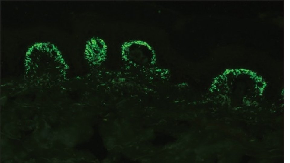
9 -Papillary Dermal Staining Granular deposits of Ig. A in the papillary dermis are diagnostic of dermatitis herpetiformis (DH). Less frequently, a similar pattern may be seen with other immunoreactants (C 3 and fibrinogen). Occasionally, a fibrillar pattern of Ig. A deposition along BMZ may be seen, especially in atypical cases of DH. Patients showing this pattern of immunoglobulin deposition may lack the circulating anti-transglutaminase and antiendomysial antibodies.
Dermatitis herpetiformis A pruritic subepidermal bullous disease associated with gluten sensitive enteropathy. The direct biopsy should be from perilesional skin because a lesional skin biopsy may be negative. DIF detects granular Ig. A deposits in the tips of dermal papillae in 80 -100% of patients. IIF shows antiendomysial Ig. A autoantibodies in 80% of patients, in subepithelial muscularis mucosa of monkey esophagus as a substrate.
10 -Exclusive Blood Vessel Wall Staining This feature is characteristically seen in cutaneous small vessel vasculitis (CSVV). The site of immune deposits in CSVV is within the walls of postcapillary venules in the superficial dermis. The most frequent immune deposits are C 3 and fibrinogen. Henoch–Schönlein purpura, a form of CSVV that is seen commonly in children, is characterized by the presence of granular staining of blood vessel wall with Ig. A with or without the presence of other immunoreactants
Vasculitis including ANCA associated small vessel vasculitides In vasculitis, DIF of skin biopsies taken from very fresh lesions display vascular deposits of Ig. M, C 3 , fibrinogen, and sometimes Ig. G. A negative result does not exclude vasculitis. Skin biopsy from HSP lesions is characterized by predominant deposition of Ig. A in the walls of upper dermal vessels. Immune deposits can be detected in skin biopsies of patients with Wegener's granulomatosis (WG) with Ig. G being the most common immunoreactant deposited in and around subepidermal blood vessels, but occasionally also along the BMZ. On IIF of sera, ANCA associated small vessel vasculitides (AASVV) are characterized by the presence of circulating ANCA. Various ELISAs directed against various ANCA specificities (including PR 3, MPO, lactoferrin, etc. ) are routinely available.
Scleroderma, dermatomyositis, and MCTDs DIF of lesional skin from patients with dermatomyositis and MCTDs may yield similar, if not indistinguishable, fluorescence results as in DLE. Nuclear keratinocyte decoration (in vivo ANA) with Ig. G and C 5 b-9 may be seen in MCTDs. In diffuse scleroderma, the frequency of positive LBT consisting of Ig. G or Ig. M and/or in vivo ANA varies from 0 to 60%.
Psoriasis It has been reported that only 3% biopsy specimens from sun-exposed psoriatic lesions may demonstrate bright continuous bands of granular positivity along the DEJ with Ig. G, Ig. M, C 3, and fibrinogen.
Cutaneous infectious diseases In infectious diseases caused by CT, HSV, and PC, the microbial agents can be easily visualized in the smears with DIF and circulating antibodies against these agents are found by IIF. DIF highlights: cytoplasmic staining in HSV type 1 -infected cells nucleolar staining in HSV type 2 -infected cells elementary bodies in CT-infected samples clusters of cysts in PC-infected samples
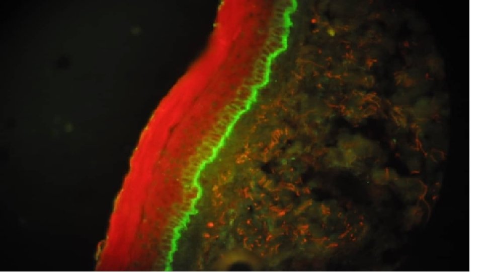
Salt-split Technique This technique was first introduced by Gammon et al. to distinguish between s. AIBDs with similar DIF findings. This technique involves artificially splitting the skin at the level of lamina lucida by incubating it in 1 M solution of sodium chloride for 24 h. There are two types of salt-split technique (SST) - direct and indirect. Direct SST employs patient's skin biopsy specimen, either freshly obtained or the one which was initially used for routine DIF. Indirect SST uses NHS as a substrate and is usually preferred over direct SST. BMZ staining in the split skin may take either a “roof” pattern (band is seen toward the epidermal side of the split) or “floor” pattern (band is seen on the dermal side of the split) or a “combined” pattern (BMZ deposits on either side of the split). In BP, antibodies tend to bind to the “roof” in 70% of the cases and in the remaining 30% of cases it reveals “combined” pattern, whereas EBA (antibodies to type VII collagen) characteristically shows a “floor” pattern on indirect SST. Other conditions that may also show “floor” pattern of staining of antibodies are antilaminin-332 subtype of MMP, anti-p 200 pemphigoid, and bullous SLE.
Salt-spit technique showing Ig. G staining on the epidermal side of the split skin in bullous pemphigoid (a) and staining with Ig. G on the dermal side in epidermolysis bullosa acquisita (b)
Pitfalls of DIF Ø It can be processed only in advanced laboratories that are proficient in the performance and interpretation of these tests. Ø It requires a thoroughly trained team comprising of a pathologist and a technologist in a specialized lab having facilities of cryostat for cutting frozen sections, − 20° C/− 80 °C deep freezers to store these sections until staining and a fluorescence microscope to report the DIF findings. Ø Skin biopsy is fragile Ø Dried and/or fixed biopsies are reported unfit for DIF. Ø The fluorescence staining quenches rapidly on exposure to light, more so under the UV light of the fluorescence microscope, resulting in the necessity of fast reporting and documentation of findings using a digital camera. Ø No long-term storage period is ideal for reporting of DIF-stained skin biopsy slides.
False-negative reactions usually occur due to technical reasons such as formalin contamination of specimen and improper transport medium or delay in shipping the sample to the laboratory. One prerequisite of IF microscopy is to keep the biopsy sample in a moist environment. Sometimes, the transport medium may leak out due to faulty closure of the lid of an aliquot, and skin biopsy reaches the laboratory in a dry state; immunoreactants in such biopsies would have undergone degradation leading onto negative result. Occasionally, biopsy sample may be devoid of epithelium - this happens when the sample is taken from the blisters or in mucosal biopsies (especially the gingival biopsy) making them suboptimal for the IF study. Recently, false-negative DIF microscopy has been reported in a case of drug-induced LAD; however, a repeat biopsy revealed the presence of linear Ig. A band in this patient. This signifies the importance of reviewing the slide, resectioning the biopsy, and if necessary repeating the biopsy in clinically suspected cases of AIBD where initial DIF is negative.
False positive results: nonspecific staining in the epidermis or BMZ may occasionally mimic the specific staining pattern leading onto diagnostic dilemma, especially to inexperienced eyes. Pemphigus-like pattern may be seen due to crushing and freezing artifacts. Nonspecific granular BMZ staining has been reported in diverse conditions such as bullous mastocytosis (with Ig. M) and elastosis perforans serpiginosa (with Ig. G). It might either be due to intense inflammation along the BMZ or nonspecific binding to the altered elastic fibers. Biopsy from the sun-exposed skin may show granular Ig. M staining along the BMZ resembling that of lupus band. Biopsy from the lower leg near the ankle may exhibit staining around the blood vessel wall, especially with fibrinogen. This may be confused with vasculitis; to avoid this, it is ideal to take biopsy from the most proximal part of the lower limb in a suspected case of vasculitis.
Advantages of IF To assist in the diagnosis of various dermatological disorders, from relatively mild ones such as LP to more lethal ones such as PV. The various circulating autoantibodies that are detected by IIF provide an indirect measure of the activity of the disease and can help in deciding which therapeutic options might be most beneficial to the patient. Antigen mapping can help in marking for a particular protein the presence or absence of which can play a role in classifying various forms of hereditary EB. The clinical picture of some infections (HSV, Chlamydia) is often muddled and unclear in patients that are immunosuppressed from any cause. In such cases, DIF can provide a rapid and convenient method to achieve a diagnosis.