Grey matter Gray matter named for its pinkishgray


- Slides: 5
Grey matter: Gray matter, named for its pinkish-gray color, is home to neural cell bodies, axon terminals, and dendrites, as well as all nerve synapses. This brain tissue is abundant in the cerebellum, cerebrum, and brain stem. It also forms a butterfly-shaped portion of the central spinal cord. Grey matter (or gray matter) is a major component of the central nervous system, consisting of neuronal cell bodies, neuropil (dendrites and unmyelinated axons), glial cells (astrocytes and oligodendrocytes), synapses, and capillaries. Grey matter is distinguished from white matter in that it contains numerous cell bodies and relatively few myelinated axons, while white matter contains relatively few cell bodies and is composed chiefly of long-range myelinated axons. The colour difference arises mainly from the whiteness of myelin. In living tissue, grey matter actually has a very light grey colour with yellowish or pinkish hues, which come from capillary blood vessels and neuronal cell bodies.
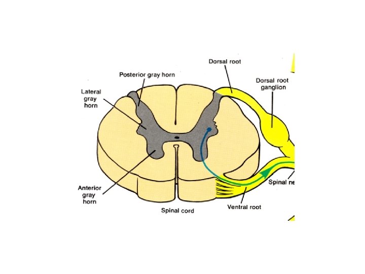
Structure: Grey matter refers to unmyelinated neurons and other cells of the central nervous system. It is present in the brain, brainstem and cerebellum, and present throughout the spinal cord. Grey matter is distributed at the surface of the cerebral hemispheres (cerebral cortex) and of the cerebellum (cerebellar cortex), as well as in the depths of the cerebrum (thalamus; hypothalamus; subthalamus, basal ganglia – putamen, globus pallidus, nucleus accumbens; septal nuclei), cerebellar (deep cerebellar nuclei – dentate nucleus, globose nucleus, emboliform nucleus, fastigial nucleus), brainstem (substantia nigra, red nucleus, olivary nuclei, cranial nerve nuclei). Grey matter in the spinal cord is known as the grey column which travels down the spinal cord distributed in three grey columns that are presented in an "H" shape. The forward-facing column is the anterior grey column, the rear-facing one is the posterior grey column and the interlinking one is the lateral grey column. The grey matter on the left and right side is connected by the grey commissure. The grey matter in the spinal cord consists of interneurons, as well as the cell bodies of projection neurons.
Grey matter undergoes development and growth throughout childhood and adolescence. Recent studies using cross-sectional neuroimaging have shown that by around the age of 8 the volume of grey matter begins to decrease. However, the density of grey matter appears to increase as a child develops into early adulthood. Males tend to exhibit grey matter of increased volume but lower density than that of females. Function: Grey matter contains most of the brain's neuronal cell bodies. The grey matter includes regions of the brain involved in muscle control, and sensory perception such as seeing and hearing, memory, emotions, speech, decision making, and self-control. The grey matter in the spinal cord is split into three grey columns: • The anterior grey column contains motor neurons. These synapse with interneurons and the axons of cells that have travelled down the pyramidal tract. These cells are responsible for the movement of muscles.
• The posterior grey column contains the points where sensory neurons synapse. These receive sensory information from the body, including fine touch, proprioception, and vibration. This information is sent from receptors of the skin, bones, and joints through sensory neurons whose cell bodies lie in the dorsal root ganglion. This information is then transmitted in axons up the spinal cord in spinal tracts, including the dorsal column-medial lemniscus tract and the spinothalamic tract. • The lateral grey column is the third column of the spinal cord. • The grey matter of the spinal cord can be divided into different layers, called Rexed laminae. These describe, in general, the purpose of the cells within the grey matter of the spinal cord at a particular location.