Central Nervous System CNS What is the function
")


 l Extracellular fluid of the Nervous System l Secreted by ependymal")


 l Protected by")

- Slides: 27
Central Nervous System (CNS)
What is the function of the CNS? 1. 2. 3. Relay messages Process information Analyze information
The Parts of the CNS 1. 2. The Brain The Spinal Cord
Cerebrospinal Fluid (CSF) l Extracellular fluid of the Nervous System l Secreted by ependymal cells l Recycled 3 x a day l Functions: l Cushions the brain l Maintain a stable fluid environment
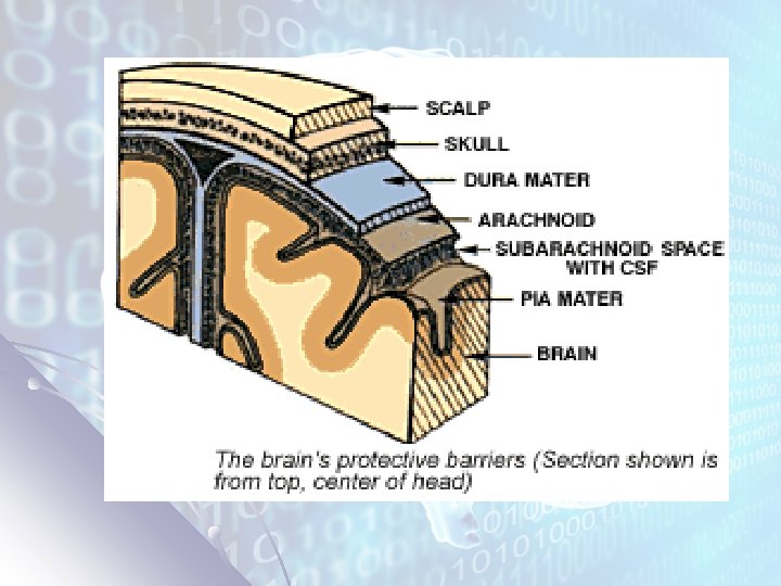
The Nervous System’s Protections l Skull and Vertebrae l 3 protective layers called meninges 1. 2. 3. l Blood-Brain Barrier l l Dura Mater (outer layer): consists of connective tissues, blood vessels and nerves. Arachnoid Layer (middle layer): elastic and web-like Pia Mater (inner layer): contains additional nerves and blood vessels. Special anatomy of CNS capillaries which limit exchange Cerebrospinal fluid l l l Separates the middle and inner layers Acts as shock absorber Exchange of nutrients between blood and nervous system
Blood-Brain Barrier
The Brain l l The brain is the control center of the body It is about 2% of your body weight BUT receives about 15% of your blood supply and uses: l ~20% of your body’s oxygen l ~50% of glucose consumed
Parts of the Brain l Divided into three main parts: 1. 2. 3. Cerebrum Cerebellum Brain Stem
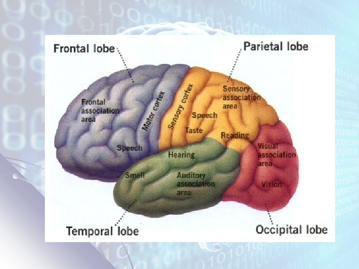
Cerebrum 1. Largest part of the brain – Site of learning and the senses l l l 2 hemispheres- Right and Left Connected by the Corpus Callosum Right side controls left side of body Left side controls right side of body Four sections or LOBES 1. Frontal Lobe 3. Parietal Lobe 2. Occipital Lobe 4. Temporal Lobe
Frontal Lobe Location: Front area of the cortex Function: Carries out higher mental processes such as thinking, decisionmaking and planning You use it to make decisions, such as what to eat for breakfast in the morning, as well as, for studying for a test. The frontal lobe is also where our personality is formed and where we can carry out higher mental processes; such as planning. In addition, the frontal lobe is necessary to being able to speak fluently and meaningfully.
Parietal Lobe Location: Upper, middle part of the cortex Function: Processes sensory information that had to do with taste, temperature and touch The parietal lobe carries out some very specific functions. As a part of the cortex, it has a lot of responsibilities and has to be able to process sensory information within seconds. The parietal lobe is where information such as taste, temperature and touch are processed. Humans would not be able to interpret sensations of touch if the parietal lobe was damaged.
Temporal Lobe Location: Bottom, middle part of cortex, right behind the temples and ears. Function: Responsible for processing auditory information from the ears (hearing) The Temporal Lobe mainly revolves around hearing and selective listening. It receives sensory information such as sounds and speech from the ears. It is also key to being able to understand speech. In fact, we would not be able to understand someone talking to us if it wasn't for the temporal lobe. This lobe is special because it makes sense of the all the different sounds being transmitted from the sensory receptors of the ears.
Location: Back part of the cortex. Occipital Lobe Function: Responsible for processing visual information from the eyes. The occipital lobe is important to being able to correctly understand what your eyes are seeing. These lobes have to be very fast to process the rapid information that our eyes are sending. Similar to how the temporal lobe makes sense of auditory information, the occipital lobe makes sense of visual information so that we are able to understand it. If our occipital lobe were injured we would not be able to correctly process visual signals, thus visual confusion would result.
Left vs Right Hemispheres
Corpus Callosum l a broad band of nerve fibers joining the two hemispheres of the brain.
Gray Matter vs. White Matter l Gray Matter – Absence of myelin in masses of neurons accounts for the gray matter of the brain – Cerebral Cortex l White Matter - Myelination gives neurons a white appearance – inner layer of cerebrum
CNS: Gray vs White Matter
CNS: Gray vs White Matter
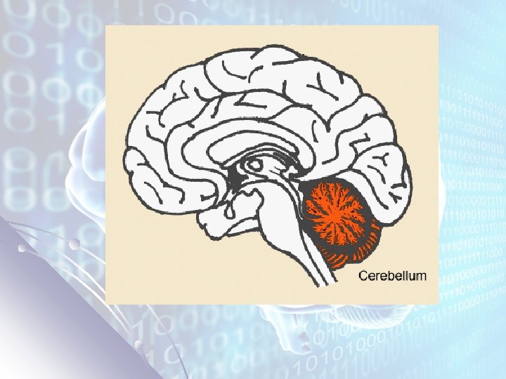
Cerebellum l l l Second largest Located below the cerebrum at back of skull This part is responsible for the balance and muscle coordination.
Brain Stem l Connects the brain to spinal cord and is made up of 2 regions: 1. Medulla Oblongata – Controls heart rate, breathing rate and flow of blood through the blood vessels. 2. Pons – Relays signals between the cerebrum and the cerebellum
Other Structures inside the Brain l Thalamus – receives messages from sensory receptors; relays information to proper regions of cerebrum l Hypothalamus - Regulates hunger, thirst, fatigue, anger, controls the pituitary gland for endocrine function
Section 35 -3 Cerebrum Thalamus Pineal gland Hypothalamus Cerebellum Pituitary gland Pons Medulla oblongata Spinal cord
Spinal Cord Link between brain and rest of body (via PNS) l Protected by the vertebrae l 31 pairs of spinal nerves (PNS) l Reflexes processed directly by spinal cord l l Reflex – quick, automatic, unconscious responses. This is a result of reflex arcs – which are the shortest nerve pathways.
Cross Section of the Spinal Cord Section 35 -3 Gray matter Spinal nerve Central canal White matter Meninges