Grand Rounds I Think I Have Worms In

- Slides: 25
Grand Rounds: “I Think I Have Worms In My Eye” Kevin Lowder, MD – PGY 3 08/24/2018 Department of Ophthalmology and Visual Sciences
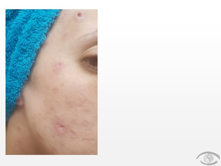
Patient Presentation CC: “I think I have worms in my eye” HPI • • 34 yo WF with PMH of non-ischemic cardiomyopathy p/w 2 -3 weeks of acneiform skin lesions on her face, out of which she has been pulling what she believes are worms. During that same time she describes a FBS in her right eye and says “I can feel a worm moving around in my right eye. ” This coincides with blurry vision OD as well. No other ocular symptoms.
Patient Presentation (continued) HPI (continued) • • No history of trauma, travel or swimming in lakes/ponds/ stagnant water, etc. No history of consuming raw/undercooked meat Pt lives on a farm where there animals, but works in an office setting Pt believes her parent’s home uses well water
Patient Presentation (continued) HPI (continued) • Multiple recent ER presentations: • • • 7/3/18 – dyspnea and syncope 7/27/18 – skin lesions on face, neck, back & chest; general malaise (since visit on 7/3/18) 8/3/18 – skin lesions, blurry vision OD, headache
History (Hx) Past Ocular Hx: none Past Medical Hx: non-ischemic cardiomyopathy, chronic pain Fam Hx: Non-contributory Meds: Tramadol, Adderall Allergies: NKDA
History (continued) Social Hx: denies Et. OH, illicit drugs* or smoking Ro. S: Skin lesions, general malaise, headaches, diarrhea
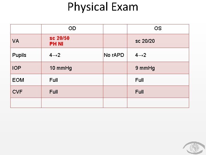
Physical Exam OD OS VA sc 20/50 PH NI Pupils 4→ 2 IOP 10 mm. Hg 9 mm. Hg EOM Full CVF Full sc 20/20 No r. APD 4→ 2
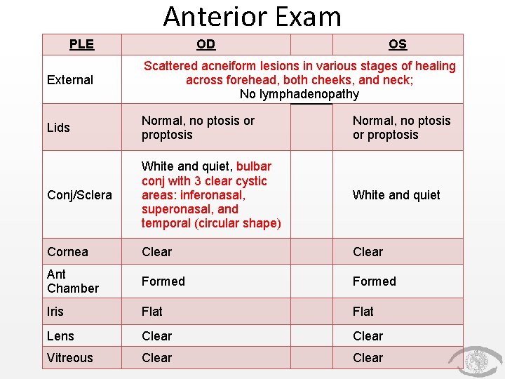
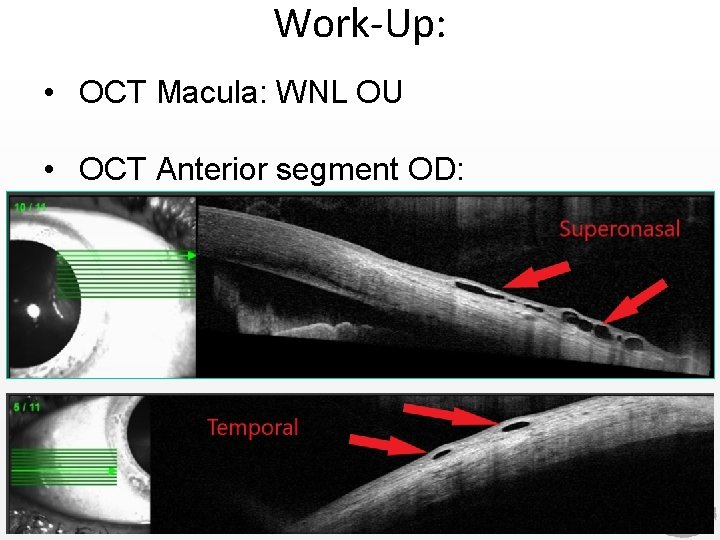
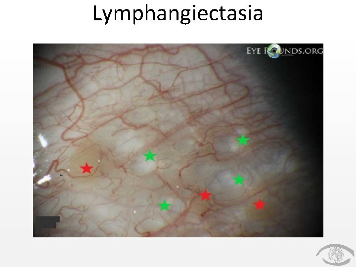
Anterior Exam PLE OD OS External Scattered acneiform lesions in various stages of healing across forehead, both cheeks, and neck; No lymphadenopathy Lids Normal, no ptosis or proptosis Conj/Sclera White and quiet, bulbar conj with 3 clear cystic areas: inferonasal, superonasal, and temporal (circular shape) White and quiet Cornea Clear Ant Chamber Formed Iris Flat Lens Clear Vitreous Clear
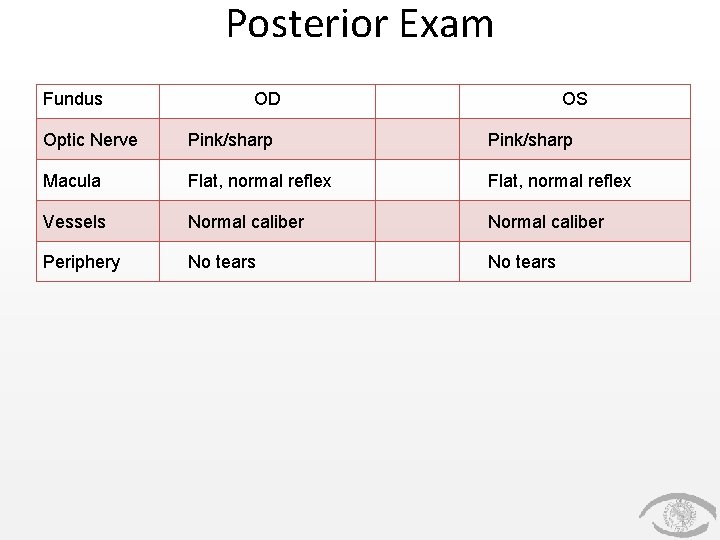
Posterior Exam Fundus OD OS Optic Nerve Pink/sharp Macula Flat, normal reflex Vessels Normal caliber Periphery No tears Normal caliber No tears
Work-Up: • OCT Macula: WNL OU • OCT Anterior segment OD:
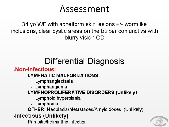
Assessment 34 yo WF with acneiform skin lesions +/- wormlike inclusions, clear cystic areas on the bulbar conjunctiva with blurry vision OD Differential Diagnosis • Non-Infectious: • • LYMPHATIC MALFORMATIONS • Lymphangiectasia • Lymphangioma LYMPHOPROLIFERATIVE DISORDERS (Unlikely) • Lymphoid hyperplasia • Lymphoma OTHER: Neoplasia/Metastases/Amyloidoses (Unlikely) Infectious (Unlikely) • Parasitic/helminthic infection
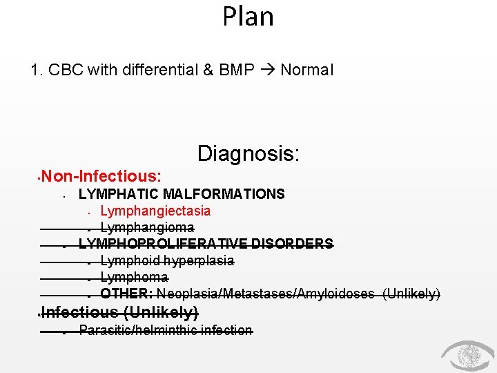
Plan 1. CBC with differential & BMP Normal Diagnosis: • Non-Infectious: • • • LYMPHATIC MALFORMATIONS • Lymphangiectasia • Lymphangioma LYMPHOPROLIFERATIVE DISORDERS • Lymphoid hyperplasia • Lymphoma • OTHER: Neoplasia/Metastases/Amyloidoses (Unlikely) Infectious (Unlikely) • Parasitic/helminthic infection
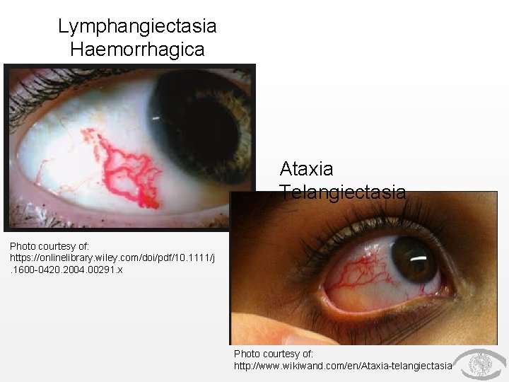
Lymphangiectasia • Irregularly dilated lymphatic channels in the bulbar conjunctiva, often associated with marked chemosis • May be developmental, or can occur following trauma or inflammation • Anomalous communication with a venule can lead to spontaneous filling with blood • *MUST DISTINGUISH THIS FROM ATAXIA-TELANGIECTASIA* • Treatment: Local excision or diathermy
Lymphangiectasia
Lymphangiectasia Haemorrhagica Ataxia Telangiectasia Photo courtesy of: https: //onlinelibrary. wiley. com/doi/pdf/10. 1111/j. 1600 -0420. 2004. 00291. x Photo courtesy of: http: //www. wikiwand. com/en/Ataxia-telangiectasia
But what about worms? ?
Discussion
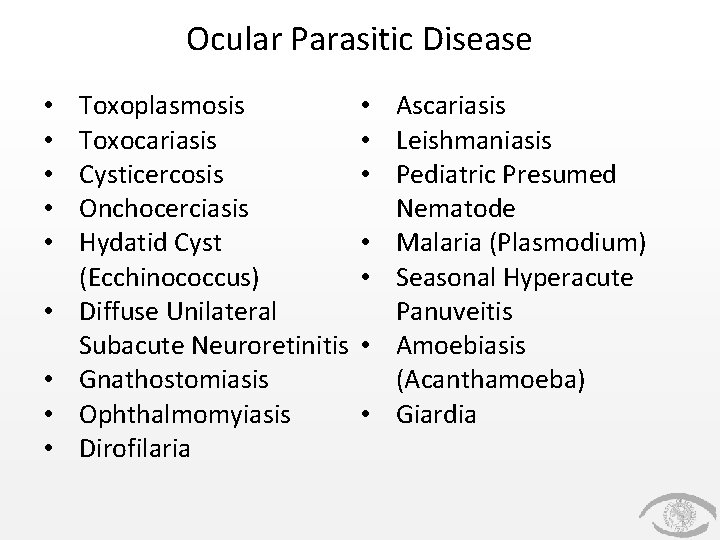
Ocular Parasitic Disease • • • Toxoplasmosis Toxocariasis Cysticercosis Onchocerciasis Hydatid Cyst (Ecchinococcus) Diffuse Unilateral Subacute Neuroretinitis Gnathostomiasis Ophthalmomyiasis Dirofilaria • Ascariasis • Leishmaniasis • Pediatric Presumed Nematode • Malaria (Plasmodium) • Seasonal Hyperacute Panuveitis • Amoebiasis (Acanthamoeba) • Giardia
Review Article – Important Points 1. Most of these infections are very uncommon (some are extremely rare) 2. Most are endemic to foreign countries/continents 3. Ocular parasitic infection leaves clues
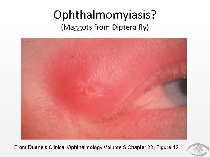
Ophthalmomyiasis? (Maggots from Diptera fly) From Duane’s Clinical Ophthalmology Volume 5 Chapter 33, Figure 42
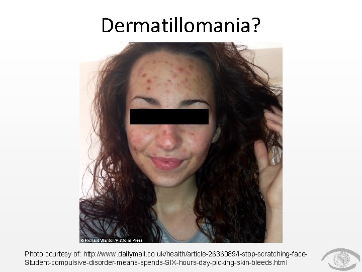
Dermatillomania? (skin picking disorder) Photo courtesy of: http: //www. dailymail. co. uk/health/article-2636089/I-stop-scratching-face. Student-compulsive-disorder-means-spends-SIX-hours-day-picking-skin-bleeds. html
Conclusions 1. Lymphangiectasia is a relatively common finding, and is often asymptomatic 2. If treatment is required, local excision or diathermy can be utilized 3. If it spontaneously bleeds from communicating with a venule, you must rule out Ataxia-Telangiectasia 4. It is quite easy to differentiate from parasitic infection
References • 2017 -2018 Basic Clinical Sciences Course, Section 8: External Disease and Cornea. American Academy of Ophthalmology, 2017. • 2017 -2018 Basic Clinical Sciences Course, Section 9: Intraocular Inflammation and Uveitis. American Academy of Ophthalmology, 2017. • https: //www. aao. org/bcscsnippetdetail. aspx? id=9 da 9385 b-8 c 6 b-466 a-8 f 9502416 a 14 f 434 • Das D, Ramachandra V, Islam S, et al. Update on pathology of ocular parasitic disease. Indian Journal of Ophthalmology. 2016; 64(11): 794 -802. doi: 10. 4103/0301 -4738. 195590.
Special Thanks *Dr. Eiferman *Dr. Scott