Endoplasmic Reticulum The endoplasmic reticulum contains a network


 • detoxification")


- Slides: 16
Endoplasmic Reticulum • The endoplasmic reticulum contains a network of branching and joining tubules 40 to 70 nanometers in diameter. • It has been calculated that 1 ml of liver tissue contains about 11 square meters of endoplasmic reticulum. • The function of the endoplasmic reticulum is to transport chemicals between and within cells. It also provides a large surface area for the organization of chemical reactions and synthesis.
Rough ER • synthesize proteins and among these are membrane proteins. • Almost all of the membrane that is created in the rough ER actually ends up providing new membrane for the ER when it is needed. • However, some of the membrane that does not get used either moves inward to replace nuclear membrane or outward to form the synthesize proteins and among these are membrane proteins. Almost all of the membrane that is created in the rough ER actually ends up providing new membrane for the ER when it is needed. However, some of the membrane that does not get used either moves inward to replace nuclear membrane or outward to form the Golgi complex, lysosomes or the plasma membrane
Smooth ER • Processes bile pigments, • glycogenolysis (the breakdown of glycogen) • detoxification of many drugs and chemical agents. • In skeletal muscles SER is involved in getting impulses from the surface of the cell into the depths of the muscle cell, which are the basis for muscle contraction.
How is the smooth endoplasmic reticulum able to detoxify drugs and poisons? The smooth endoplasmic reticulum is richly concentrated in enzymes that specialize in making chemical modifications to drugs, particularly in liver cells. Oxidases are important in this role because they specialize in oxidizing, in most cases adding oxygen atoms to drug molecules. For drugs to stay in the body; they are not very soluble in water and therefore are not passed through the urine. Even when drugs travel through the bloodstream, they remain bound to plasma proteins and other things in the blood and thus remain invisible to the kidney.
• The enzymes in the smooth endoplasmic reticulum of the liver try to make drugs more soluble in water. One way to do this is to make them more "polar, " in other words, to add electronegative atoms such as oxygen to the structure. In many cases, the same modifications that make a drug more water soluble also eliminate its intended effect. Therefore, they are being handled by liver enzymes, the drug molecules no longer work.
Golgi Apparatus • The Golgi apparatus, also called Golgi body or Golgi complex and found universally in both plant and animal cells, is typically comprised of a series of five to eight cup-shaped, membrane-covered sacs called cisternae that look something like a stack of deflated balloons.
• Is often considered the distribution and shipping department for the cell's chemical products. • It modifies proteins and lipids (fats) that have been built in the endoplasmic reticulum • prepares them for export outside of the cell or for transport to other locations within the cell
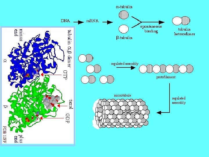
Microtubules • Are filamentous intracellular structures that are responsible for various kinds of movements in all eukaryotic cells. • Are involved in nucleic and cell division • organization of intracellular structure, • intracellular transport, • ciliary and flagellar motility.
Specialty Microscopes • http: //micro. magnet. fsu. edu/primer/java/ele ctronmicroscopy/magnify 1/index. html
Transmission Electron Microscope TEM • The TEM was developed as an optical instrument, for imaging specimen structures at high resolution beyond that possible in an optical microscope. The first instruments were developed in the 1930's. Electrons are useful in the formation of images because they have an extremely short wavelength which is dependent on their energy.
TEM
Scanning Electron Microscope • Unlike specimens for transmission microscopy, the SEM specimen does not have to be thin enough for electrons to travel through, and SEMs are routinely used to look at the surfaces of bulk specimens. Although the first description of an SEM appeared in 1935, it was not until the 1960's that the first commercial instruments appeared.
SEM cheese mold spore chains
Scanning Tunneling Microscopy • The invention of the scanning tunneling electron microscope in 1981 allowed scientists to start manipulating atoms and molecules, pushing them into interesting shapes. They demonstrated this new ability by making a molecular abacus and a molecular wheel.
These are atoms