Department of faculty and hospital surgery Tashkent Medical


 began thoracic surgery. The first")
 The clinic as a diagnostic method thoracoscopy")

 Boris Petrovsky (1908 - 2004) Ivan Greeks (1867 -1934) Mikhail")


- Slides: 44
Department of faculty and hospital surgery Tashkent Medical Academy Pleural diseases
The prevalence of pleural diseases n n Empyema refers to the common diseases of the chest. In 30 -50% of cases, it complicates the course of acute and chronic pneumonia, 80 -90% - of gangrene of the lung, in the 9 -15% - lung abscess, 6 -8% - bronchiectasis At 4 -34 patients with acute empyema developed after surgical treatment for lung and pleura. In 1. 3 -3 times more disease in men
With the treatment of diseases of the pleura (empyema) began thoracic surgery. The first description of thoracic surgery belongs to Hippocrates, who diagnosed pleural empyema and cured the sick releasing the pus by a cut (now it's called torakostomiya) Hippocrates (about 460 years BC)
n n Hans Christian Yakobeus (1879 -1937) The clinic as a diagnostic method thoracoscopy was first performed by the Swedish physician, pulmonologist Hans Christian Yakobeusom in 1910 with the help of a cystoscope to diagnose the state of the pleural cavity in patients with tuberculosis. Later Yakobeus designed optical instrument called thoracoscope, whereby initially inspect the pleural cavity, and from 1910 to 1913, he completed 89 of thoracoscopy. In 1913 Yakobeus upgraded Thoracoscope, adjoining the galvanokauter, and began using thoracoscopy for pleural adhesions perezhiganiya. In 1925, he first performed pleural biopsy in patients with mesothelioma and reported on the implementation of 120 thoracoscopy in patients with pulmonary tuberculosis.
In 1925, PA Herzen performed the first in the USSR thoracoscopy in chronic pleural empyema. Subsequently, it is most often used in TB for the intersection of adhesions by galvano-cautery for the formation of a therapeutic pneumothorax. n Peter A. Herzen (1871 -1947)
Sergey Yudin (1891 -1954) Boris Petrovsky (1908 - 2004) Ivan Greeks (1867 -1934) Mikhail Davydov
Vasit Vakhidovich Vahidov Shavkat Ibrahimovic Karimov Sadik Aliyevich Masumov Nikolai Fedorovich Krotov
Duties of the general practitioner in diseases of pleura n n n n n - Provision of primary health and social care; - Health education (promoting healthy lifestyles); - Preventive work (timely detection of early and latent forms of the disease, risk groups); - Dynamic monitoring; - Emergency assistance in case of emergency and acute conditions; - Timely consultation and hospitalization in the prescribed manner; - Medical and rehabilitation work in accordance with the qualifying characteristic; - An examination of temporary disability; - The organization of medical and social care and household together with the bodies of social protection and services of mercy alone, the elderly, the disabled, the chronically ill; - Maintaining the approved forms of records and reports.
Anatomy of the pleura
Anatomy of the parietal pleura 1. Neck part 2. Costal part 3. Mediastinal part 4. Diaphragmatic part
Classification of Diseases of the pleura Empyema 2. Spontaneous pneumothorax 3. Injuries of the chest 4. Clotted hemothorax 5. Fibrothorax 6. Tumor of the pleura 1.
Classification of empyema I. By the origin primary secondary II. The clinical course acute (disease lasts less than 8 weeks) chronic (illness lasts more than 8 weeks) III. By the nature of exudate purulent putrid IV. By the nature of the microflora specific (tuberculosis, fungal) nonspecific (staphylococcal, dyplococcal, anaerobic) mixed V. By the distribution process total and subtotal limited empyema: parietal, basal, interlobarnaya, apical, mediastinal an multi-chamber VI. Iatrogenic empyema
Clinical signs of pleural empyema n n n Cough Sputum Signs of intoxication Fever Asymmetry of chest Auscultation reduction or absence of pulmonary respiration in the affected side
Empyema
Empyema
Empyema radiography computed tomography
Changing the location of the fluid in the pleural cavity according to the position of the patient
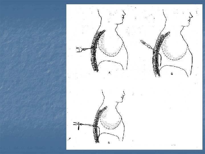
Puncture of the pleural cavity
The location of the needle with respect to the fins during the puncture of the pleural cavity
Thoracostomy with the help of the trocar
Technique of chest draining
The valve is used for drainage of the pleural cavity
Device for drainage of the pleural cavity by Byulau
Thoracostomy by Bobrov
Modern plant for active aspiration of the pleural cavity
The scheme of pleuroevacuator
Pleuroevacuator
Methods of rehabilitation of the pleural cavity
Decortication of the lung
As a rule, spontaneous pneumothorax is a complication, not a cause
Spontaneous pneumothorax
Position of the patient on the operating table Intubation tube to separate light
Thoracoscopic intervention in spontaneous pneumothorax Coagulation of the bull Ligation of the bull Suturing of the bull Partial atypical resection Diagnostic thoracoscopy
The advantages of VATS intervention n n n n Reducing trauma surgery Reduction in pain There is no need to find a patient in the intensive care unit Early activation of patients Reducing the number of complications associated with physical inactivity on the part of the surgical wound Reduction in length of hospital stay Reduction in general terms of disability Reducing the flow of drugs and surgical material Good cosmetic effect
Pronounced cosmetic effect after thoracoscopic interventions
Radiographic signs of damage to the chest n n n n n Partial or complete alveolar infiltrates Mediastinal shift Subcutaneous emphysema Pneumomediastinum Fractured ribs Expansion of the shadow of the mediastinum The disappearance of the contour of the aortic arch Mixing of a nasogastric tube to the right Expansion of cardiac contour Presence of gastric bubble and bowel loops in the pleural cavity
Clinical signs of damage at the chest 1. 2. 3. 4. 5. 6. 7. Cyanosis The absence of spontaneous breathing Subcutaneous emphysema The presence of abnormal noise in the pleural cavity The presence of chest injury Bulging neck veins Paradoxical pulse
Hemothorax
Tension pneumothorax Subcutaneous emphysema n For percussion tympanitis on the affected side n Mixing of the trachea in a healthy way n The weakening of the respiratory noise n Unstable hemodynamics n
Open pneumothorax Tension pneumothorax schematic representation of
Treatment of injuries of the chest 1. 2. 3. Thoracostomy Surgical treatment Conservative therapy
Indications for drainage of the pleural cavity 1. 2. 3. Pneumothorax Injuries of the chest Hemothorax
Indications for surgical treatment of injuries of the chest n n n n Cardiac tamponade Large open wound of the chest Signs of damage to the mediastinum Profuse bleeding continues or pleural cavity Massive pnevmoreya on drainage of the pleural cavity Damage to the trachea or main bronchus Rupture diaphragms, aorta, esophagus Foreign body chest