Cerebral Cortex Cerebellum The cerebrum n The largest

Cerebral Cortex & Cerebellum

The cerebrum n The largest portion of the brain. n The surface is marked by ridges called Gyri separated by: Fissures ► 2 hemispheres connected by the corpus callosum. Ø Sulci ► lobe The cerebral hemispheres consist of a convoluted cortex of grey matter overlying the central medullary mass of white matter. The grey matter of the cerebral cortex has a very extensive surface area provided by the convoluted gyri separated by sulci and fissures. The medulla of the cerebrum conveys fibers between different parts of the cortex to and from other parts of the CNS. The white matter of the cerebral hemispheres contains collection of nerve cells called the basal ganglia.

The grey matter It is called grey, because its color in the fresh state is grey. It is composed of: A complex of nerve cell bodies. Nerve fibers (mostly unmyelinated) A network of neuroglia A rich capillary bed. The white matter It is called white because its color in the fresh state is white. It is composed of: Bundles of nerve fibers (mostly myelinated) A network of neuroglia Few blood capillaries.

Histological examination of a section of the cerebral cortex: 1 - cell bodies of neurons ► ► laminae 2 - nuclei of supporting neuroglia. 3 - capillaries. 4 - The neuropil = meshwork ► ► sensitive to changes in O 2 & sugar. H&E: structureless substance

EM: network of 1. Nerve cell processes. 2. Cell bodies and processes of astrocytes. 3. Intercellular spaces containing tissue fluid
 2")
Types of neurons in the cerebral cortex They are arranged in layers (laminae) 2 main types 1. Pyramidal cells: 2. Stellate cells: A wide variety of stellate cells are present in the cortex: Horizontal cells of Cajal: are small, spindleshaped and oriented parallel to the surface. Fusiform cells: They are spindle-shaped cells oriented at right angles to the surface of cerebral cortex. Granule cells: Granule cells are present in all cortical laminae. Cells of Martinotti: They are small cells with long axon which extends towards the surface
")
6 Layers of the cerebral cortex in the motor area 1 - Molecular layer(plexiform) ▲Fibers: parallel to surface. = apical dendrites of pyramidal + axons of granule & Martinotti cells. ▼Cell bodies: horizontal cells of Cajal Neuroglia 2 - External granular layer Cell bodies: ▲ ▲ granule cells 3 - External pyramidal cell layer Cell bodies: pyramidal: small & med-sized

4 - Internal granular layer Cell bodies: ▲ ▲ granule cells 5 - Internal pyramidal cell layer Cell bodies: med. - sized & Large pyramidal cells Betz cells 6 - The polymorphic layer multiform The deepest & broadest Cell bodies: cells of Martinotti

Cyto-architecture of some cerebral areas The cerebral cortex shows the same general structure (laminar pattern) with certain modifications in some cortical areas to perform different functions. A Brodmann area is a region of the cerebral cortex , defined by its cytoarchitecture , or histological structure and organization of cells 1. The motor area. It is of the agranular cytological type. It has few scattered granule cells, while the pyramidal cell layers are well developed. Betz cells are found in the inner pyramidal layer. 2. The sensory area It is of granular type. The granular layers are well developed, whereas, the pyramidal layers

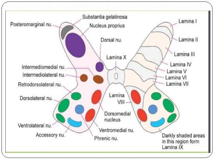
Rexed laminae The Rexed laminae are layers of neurons within the spinal cord that perform specific functions. comprise a system of ten layers of grey matter (I–X) Laminae Posterior grey column : I–VI Lamina I: marginal nucleus of spinal cord or posteromarginal nucleus Lamina II: substantia gelatinosa of Rolando Laminae III and IV: nucleus proprius Lamina V: Neck of the dorsal horn. Neurons within lamina V are mainly involved in processing sensory afferent stimuli from cutaneous, muscle and joint mechanical nociceptors as well as visceral nociceptors. Lamina VI: Base of the dorsal horn.

Lateral grey column : VII and X Lamina VII: intermediomedial nucleus, intermediolateral nucleus , posterior thoracic nucleus in the thoracic and upper lumbar region Lamina X: an area of grey matter surrounding the central canal Anterior grey column : VIII–IX Lamina VIII: motor interneurons ; Commissural nucleus Lamina IX: body wall muscles , lateral (in limb regions) and medial (back muscles) motor neurons , also phrenic and spinal accessory nuclei at cervical levels, and nucleus in the sacral region

Histology of the cerebellum

The Cerebellum Lies in the posterior cranial fossa covered by tentorial membrane. 2 nd largest region of the brain. 10% of the brain by volume, but it contains 50% of its neurons. separated from the brain stem by 4 th ventricle. 2 hemispheres separated by the vermis. Its surface is marked by T. S curved fissures (laminated appearance).

The Cerebellar cortex • forms Folia, a branching array that in a sectional view resembles a tree • External grey matter + core of white matter • The deep cerebellar nuclei: 1 -dentate: in the median plane. 2 - emboliform. 3 - globose.

Layers of the cerebellar cortex 1 -Molecular layer: 2 - Purkinje cell layer. 3 - Granular layer. 1 -Molecular layer: ▲▲Fibers: - dendrites of Purkinje cells + Golgi cells. - climbing fibers= ( from inferior olive to Purkinje cells) - axons of granular cells. ▼▼Cells: - basket cells - neuroglia.

2 - Purkinje cell layer: layer huge pyriform, in single layer whose extensive dendritic arborization Axons. . . . to deep cerebellar nuclei.

3 - granular layer: • Densely packed small stellate granule cells: • receive mossy fibers. • Axons to molecular layer. • Golgi cells: cells • extensive branching dendrites in different planes. . . • receive recurrent collateral fibers. G P

The fiber architecture of the cerebellar cortex: It includes: 1 - Afferent fibers are the fiber inputs to the cerebellar cortex, these are: Mossy fibers which constitute the majority of afferent fibers to the cerebellar cortex. They end on the granule cells. Climbing fibers which are the olivo- cerebellar fibers that end on the dendrites of Purkinje cells. Recurrent collaterals arise from axons of Purkinje cells and end on dendrites of Golgi cells. 2 -Efferent fibers are the axons of Purkinje cells that end on the deep cerebellar nuclei. RC C M E

The cerebellar glomeruli: These are the complex synaptic regions between: • mossy fibers (Rosettes endings)+ • dendrites of granule cells • axons of Golgi cells. • enclosed in neuroglial capsule. characterize the granular layer.

The Brain Barriers The neural tissue of the brain and spinal cord are protected by several barriers which preserve the homeostasis (internal environment) of the CNS. These are: 1 - Blood- CSF barrier: It keeps the chemical stability of the CSF different from that of the blood plasma. This barrier is situated at the tight junctions between the ependymal cells of the choroid plexus. 2 - Brain- CSF barrier: It separates the CSF from the surrounding brain tissue. It is formed of: The ependymal cells lining the brain ventricles and the central canal of the spinal cord.

3 - Blood- brain barrier: It is a highly selective barrier that prevents the passage of harmful materials e. g. toxins, foreign proteins and some drugs. In addition, it allows the exchange of gases (O 2, CO 2) and many simple solutions (glucose) essential for nutrition of neurons. The blood- brain barrier is established by : I- The specific microscopic structure of CNS capillaries which is formed of: • The non-fenestrated capillaries of the CNS with tight (occluding) junctions between the lining endothelial cells. • The CNS capillaries are invested by continuous basal lamina. • Neuroglial processes (end feet of astrocytes) that completely surround the blood capillaries. II- The physiological transport system: that controls the endothelial permeability of blood- born substances. The trans- endothelial transport of CNS capillaries is completely restricted to receptor- mediated transport with very few pinocytotic vesicles.

CSF circulation
- Slides: 24