Cell Fractionation and Ultracentrifugation Cell Fractionation What is


- Slides: 9
Cell Fractionation and Ultracentrifugation
Cell Fractionation What is it? • Process where cells are broken up and the different organelles they contain separated out. • Used to obtain large numbers of isolated organelles • Used to help us study structure and function of organelles
Cell Fractionation Before cell fractionation can begin tissue placed in cold, isotonic buffer solution… -Cold to reduce enzyme activity which may break down organelles -Isotonic (same water potential as original tissue) to prevent organelles bursting/shrinking due to osmotic gain/loss of water. -Buffered to maintain a constant p. H so that structure of organelles is not altered and enzyme activity is unaltered Two-stage process: homogenation and ultracentrifugation.
Steps of Cell Fractionation & Ultra Centrifugation 1. Tissue to be studied is cut into small pieces (Minced) and placed into a COLD, ISOTONIC and BUFFERED solution. 2. These are then ground into smaller pieces using a Homogenizer (Sophisticated liquidizer) releases the organelles from the cell. 3. The Homogenate is filtered to remove any complete cells and large debris i. e. Cell Wall/Membrane 4. A suspension of homogenate is then placed in a test tube and then centrifuged – The faster the speed at which the tube is spun, the greater the force generated. 5. At slower speeds the larger fragments collect at the bottom of the tube and smaller ones remain near the top suspended in a liquid called the SUPERNATENT LIQUID. 6. These larger fragments (Sediment Pellets) are then removed and the supernatant remaining is re-spun at a faster speed (more force) and some of the smaller fragments collect at the bottom forming a new pellet. 7. By continuing in this way smaller and smaller fragments will be recovered.
• The size of any organelle is relatively constant so separate at a specific speed • Since the whole process involves centrifuging at different speeds it is called DIFFERENTIAL CENTRIFUGATION Order of organelle fractionation Speed of centrifugation x Gravity Duration of spin (mins) Organelle Pellet formed 1000 10 Nuclei 3, 500 10 Chloroplasts Mitochondria 16, 500 20 Lysosomes Endoplasmic Reticulum 100, 000 60 Ribosomes
Cell Fractionation Video Clips https: //www. youtube. com/watch? v=SCNEf. R 85 a. Yk (11 mins) https: //www. youtube. com/watch? v=Sja. FUJhz. Y 9 Q (2. 2 mins)
Task: Create a flow chart showing the process of cell fractionation using the steps below. Add detail using pages 59 -60 Tissue is homogenised e. g. in a blender Supernatant is analysed for content. Homogenised suspension is filtered Tissue is cut into pieces in cold, buffered, isotonic solution. Supernatant is decanted and re-centrifuged at higher speeds until desired organelle is separated. Filtrate is centrifuged at low speed
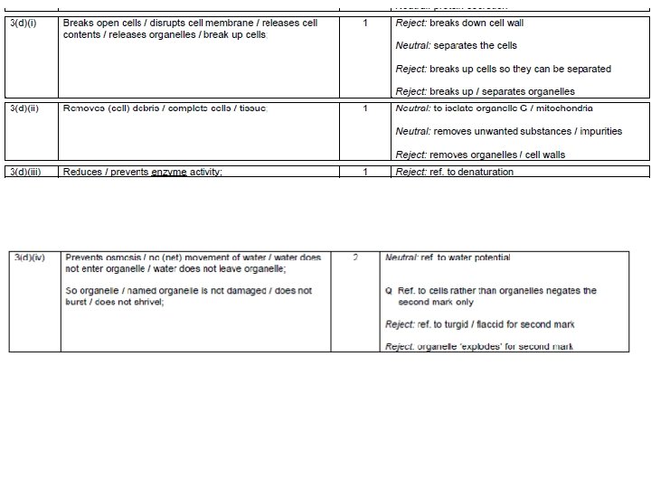
Exam Question.