ATILLA ERTAN MD FACP AGAF MACG F G


- Slides: 29
ATILLA ERTAN, MD, FACP, AGAF, MACG
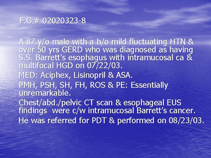
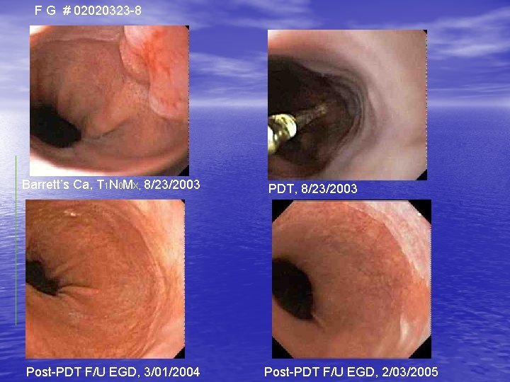
F. G. # 02020323 -8 A 87 y/o male with a h/o mild fluctuating HTN & over 50 yrs GERD who was diagnosed as having S. S. Barrett’s esophagus with intramucosal ca & multifocal HGD on 07/22/03. MED: Aciphex, Lisinopril & ASA. PMH, PSH, FH, ROS & PE: Essentially unremarkable. Chest/abd. /pelvic CT scan & esophageal EUS findings were c/w intramucosal Barrett’s cancer. He was referred for PDT & performed on 08/23/03.
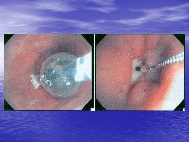
F G # 02020323 -8 Barrett’s Ca, T 1 N 0 MX, 8/23/2003 Post-PDT F/U EGD, 3/01/2004 PDT, 8/23/2003 Post-PDT F/U EGD, 2/03/2005
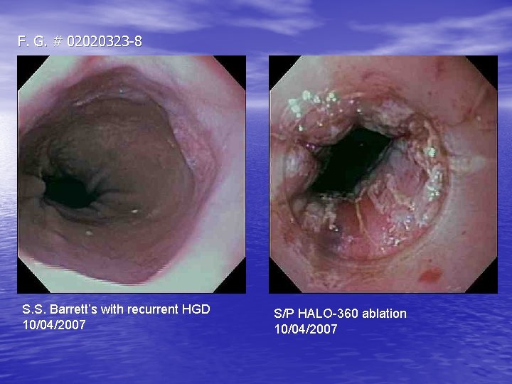
F. G. # 02020323 -8 S. S. Barrett’s with recurrent HGD 10/04/2007 S/P HALO-360 ablation 10/04/2007
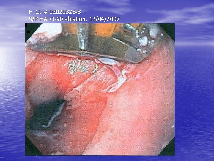
F. G. # 02020323 -8 S/P HALO-90 ablation, 12/04/2007
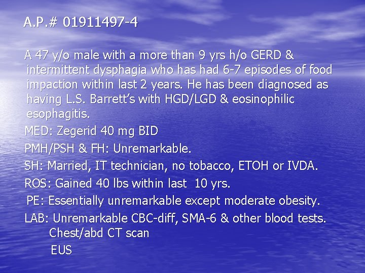
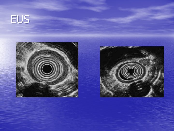
A. P. # 01911497 -4 A 47 y/o male with a more than 9 yrs h/o GERD & intermittent dysphagia who has had 6 -7 episodes of food impaction within last 2 years. He has been diagnosed as having L. S. Barrett’s with HGD/LGD & eosinophilic esophagitis. MED: Zegerid 40 mg BID PMH/PSH & FH: Unremarkable. SH: Married, IT technician, no tobacco, ETOH or IVDA. ROS: Gained 40 lbs within last 10 yrs. PE: Essentially unremarkable except moderate obesity. LAB: Unremarkable CBC-diff, SMA-6 & other blood tests. Chest/abd CT scan EUS
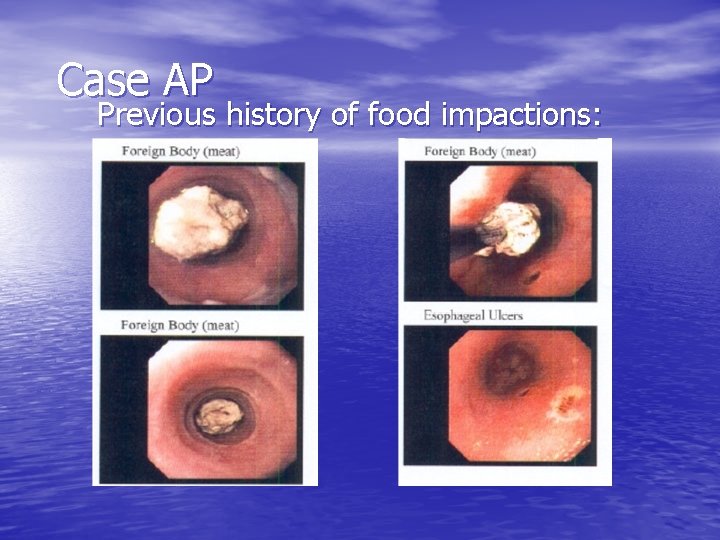
Case AP Previous history of food impactions:
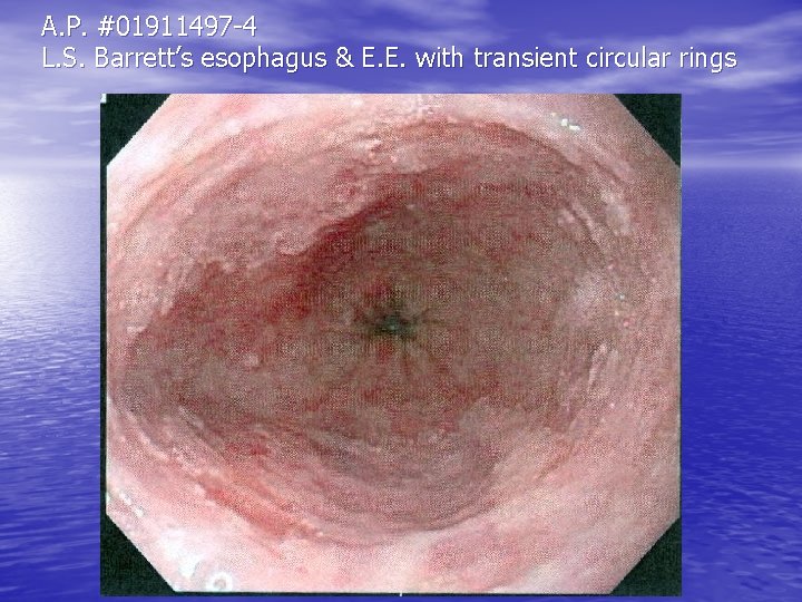
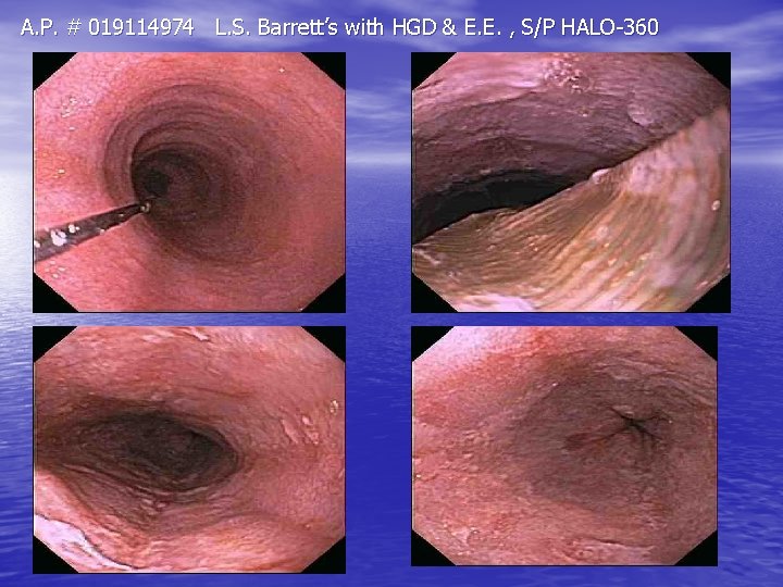
A. P. #01911497 -4 L. S. Barrett’s esophagus & E. E. with transient circular rings
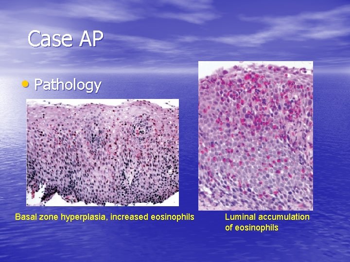
Case AP • Pathology Basal zone hyperplasia, increased eosinophils Luminal accumulation of eosinophils
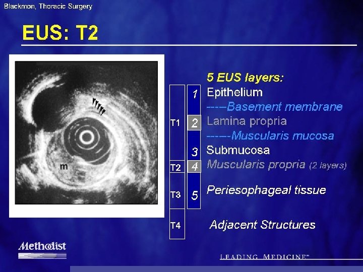
EUS
What would you do next? • Stage: • T 3 N 0 M 0 Esophageal Cancer • GEJxn Type II Tumor
A. P. # 019114974 L. S. Barrett’s with HGD & E. E. , S/P HALO-360
EOSINOPHILIC ESOPHAGITIS [EE] & BARRETT’S ESOPHAGUS • “Barrett’s esophagus or esophageal adenocarcinoma has not been reported in patients with EE” (1, 2). • “EE is not a disease characterized by mucosal ulceration or destruction. Therefore, it seems likely that the pathologic process of EE is different from that of GERD and that adenocarcinoma or squamous cancer of the esophagus are not the spectrum of EE, other than perhaps as coincidental occurences” (2). Natural history & long-term follow-up studies are needed to provide more information in this relation. 1. Am J Gastroenterol, 101: 1900, 2006. 2. Gastroenterology, 133: 1342, 2007.
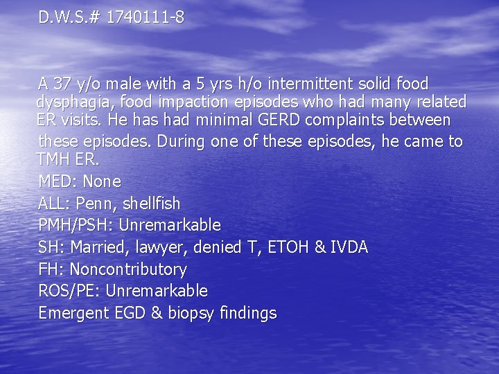
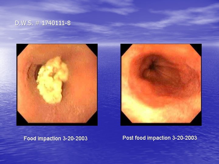
D. W. S. # 1740111 -8 A 37 y/o male with a 5 yrs h/o intermittent solid food dysphagia, food impaction episodes who had many related ER visits. He has had minimal GERD complaints between these episodes. During one of these episodes, he came to TMH ER. MED: None ALL: Penn, shellfish PMH/PSH: Unremarkable SH: Married, lawyer, denied T, ETOH & IVDA FH: Noncontributory ROS/PE: Unremarkable Emergent EGD & biopsy findings
D. W. S. # 1740111 -8 Food impaction 3 -20 -2003 Post food impaction 3 -20 -2003
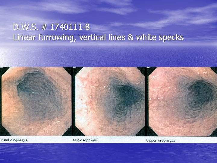
D. W. S. # 1740111 -8 Linear furrowing, vertical lines & white specks
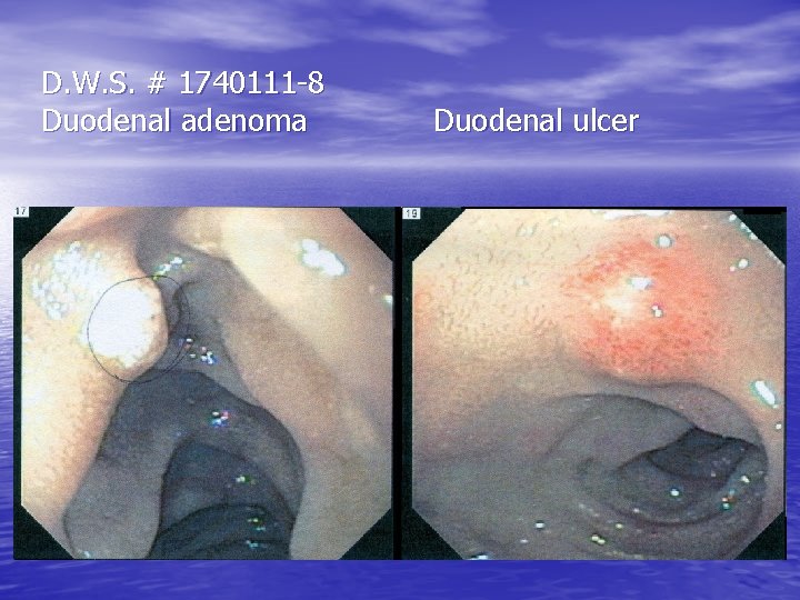
D. W. S. # 1740111 -8 Duodenal adenoma Duodenal ulcer
EOSINOPHILIC ESOPHAGITIS [EE] __________________________ During the last decade, we saw a rapid increase of patients with esophageal intraepithelial eosinophilia who were thought to be GERD but who did not respond to GERD management. Subsequent studies showed that these patients had a “new “ disease termed EE which is a disease characterized by: • Symptoms including but not restricted to food impaction & dysphagia in adults , and feeding intolerance & GERD symptoms in children. • ≥ 15 intraepithelial eosinophilis/HPF • Exclusion of other disorders associated with similar clinical, histological, or endoscopic features, especially GERD. __________________________ FIGERS; Gastroenterol 133: 1342 -63, 2007.
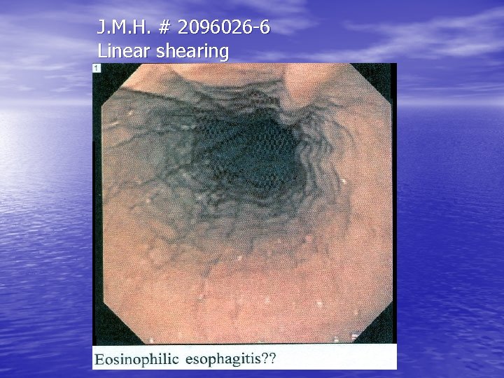
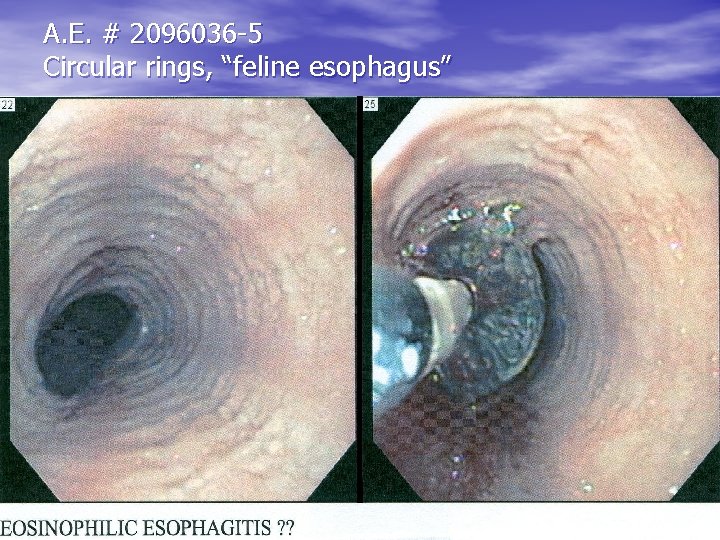
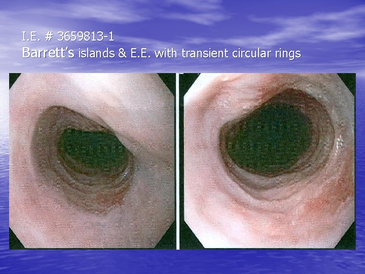
ENDOSCOPIC FEATURES ASSOCIATED WITH E. E. ___________________ • Unremarkable endoscopic mucosa & lumen. • Circular rings, transient or fixed, “feline esophagus” • Linear furrowing, vertical lines of the mucosa • Linear shearing/crepe paper mucosa with passage • • of endoscope or dilator White exudates, white specks, nodules or granularity Stricture/rings: proximal, middle, or distal. ___________________ FIGERS: Gastroenterol, 133: 1342, 2007 (modified). * None of the features are pathogonomic of EE.
J. M. H. # 2096026 -6 Linear shearing
A. E. # 2096036 -5 Circular rings, “feline esophagus”
I. E. # 3659813 -1 Barrett’s islands & E. E. with transient circular rings
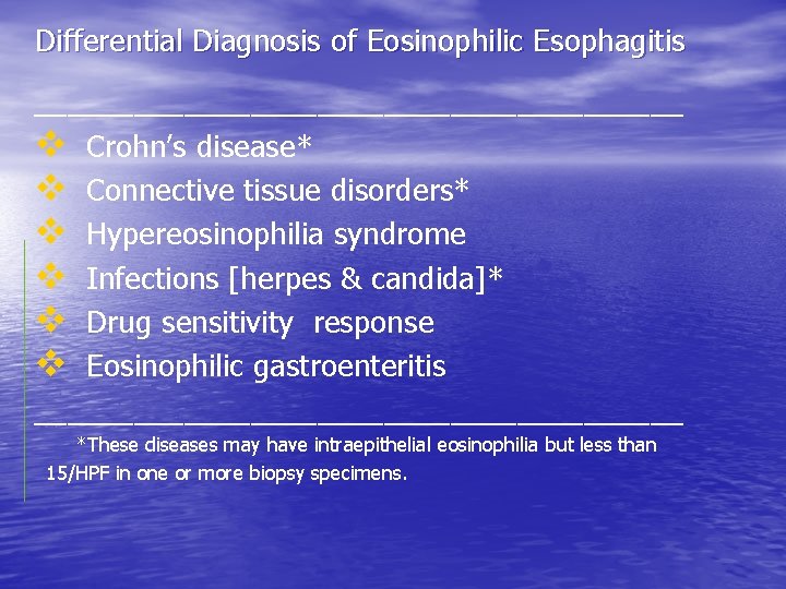
Differential Diagnosis of Eosinophilic Esophagitis ____________________ v Crohn’s disease* v Connective tissue disorders* v Hypereosinophilia syndrome v Infections [herpes & candida]* v Drug sensitivity response v Eosinophilic gastroenteritis ____________________ *These diseases may have intraepithelial eosinophilia but less than 15/HPF in one or more biopsy specimens.
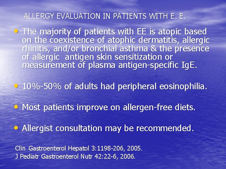
ALLERGY EVALUATION IN PATIENTS WITH E. E. • The majority of patients with EE is atopic based on the coexistence of atophic dermatitis, allergic rhinitis, and/or bronchial asthma & the presence of allergic antigen skin sensitization or measurement of plasma antigen-specific Ig. E. • 10%-50% of adults had peripheral eosinophilia. • Most patients improve on allergen-free diets. • Allergist consultation may be recommended. Clin Gastroenterol Hepatol 3: 1198 -206, 2005. J Pediatr Gastroenterol Nutr 42: 22 -6, 2006.
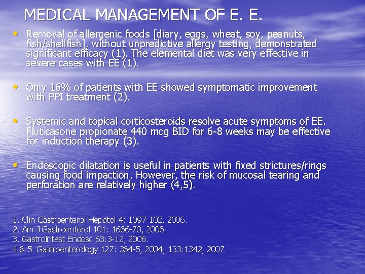
MEDICAL MANAGEMENT OF E. E. • Removal of allergenic foods [diary, eggs, wheat, soy, peanuts, fish/shellfish], without unpredictive allergy testing, demonstrated significant efficacy (1). The elemental diet was very effective in severe cases with EE (1). • Only 16% of patients with EE showed symptomatic improvement with PPI treatment (2). • Systemic and topical corticosteroids resolve acute symptoms of EE. Fluticasone propionate 440 mcg BID for 6 -8 weeks may be effective for induction therapy (3). • Endoscopic dilatation is useful in patients with fixed strictures/rings causing food impaction. However, the risk of mucosal tearing and perforation are relatively higher (4, 5). 1. Clin Gastroenterol Hepatol 4: 1097 -102, 2006. 2. Am J Gastroenterol 101: 1666 -70, 2006. 3. Gastrointest Endosc 63: 3 -12, 2006. 4 & 5. Gastroenterology 127: 364 -5, 2004; 133: 1342, 2007.