Thoracic Volume Modeling in Early Onset Scoliosis David


 increases, thoracic")


- Slides: 15
Thoracic Volume Modeling in Early Onset Scoliosis David Matson, MS 21; Charles Ledonio, MD 2; David Polly Jr. , MD 2; Kristin England, MD 2; Jeff Pawelek, BS 4; and Behrooz Akbarnia, MD 3, 4 1 University of Minnesota Medical School; Minneapolis, MN. 2 Department of Orthopaedic Surgery, University of Minnesota Medical School; Minneapolis, MN. 3 Department of Orthopaedic Surgery, University of California, San Diego; San Diego, CA. 4 San Diego Center for Spinal Disorders; La Jolla, CA.
Disclosures • David Matson: no disclosures • Charles Ledonio, MD: – Grants/Research: Medtronic, Do. D, OREF • David Polly, Jr, MD: – Grants/Research: Do. D, OREF • Kristin England, MD: no disclosures • Jeffery Pawelek: no disclosures • Behrooz Akbarnia, MD: – – Grants/Research: De. Puy Spine Consulting: Ellipse, Kspine, K 2 M Ownership/Shareholder: Nuvasive, Ellipse, Kspine Royalty/Patent: De. Puy Spine, K 2 M
Background • Virtual thoracic volume modeling from plain radiographs has been used in the adolescent idiopathic scoliosis (AIS) and early onset scoliosis (EOS) populations. Thoracic volume from modeling correlates within 3 -4% of thoracic volume from CT scans. • Early onset scoliosis (EOS) ↓ thoracic volume and lung volume • For AIS patients with poor pulmonary function, the modeled 2 year post-op thoracic volume change is strongly correlated with the two year post-op pulmonary function test.
Virtual modeling of scoliotic deformity • • As coronal deformity (Cobb Angle) increases, thoracic volume decreases Cobb Angle >70⁰, sagittal deformity does not appear to impact thoracic volume (England)
Purpose • Objective: to assess thoracic volume change with growing rod interventions in patients with early onset scoliosis.
Methods • • • Retrospective case study of children 10 years of age and younger with diagnosis of EOS Convenience sample of 6 patients with EOS from Growing Spine Study Group Coronal and sagittal radiographs used to model thoracic volume
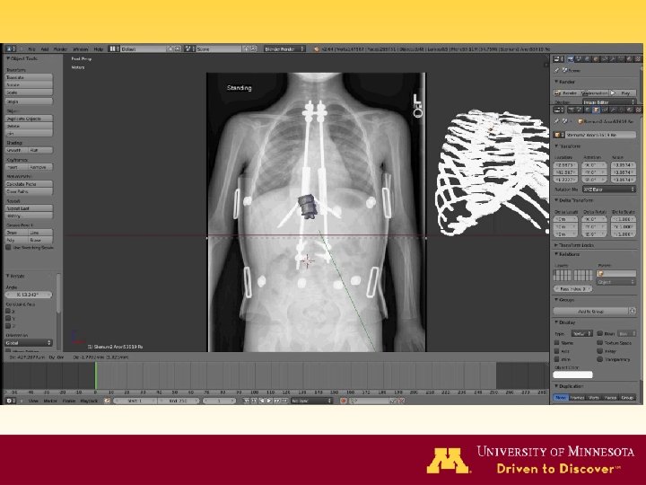
Methods • • Patients all underwent growing rod surgery with varying number of lengthening procedures for treatment of diagnosed early onset scoliosis Blender software (2. 71, open access) to create 3 D model from coronal and sagittal radiographs – ‘computationally deformed’ to match chest X-rays • 3 D models created with pre- and post-operative radiographs – Post-op taken from midpoint of treatment and final lengthening – Up to 3 models per patient • Thoracic volume determined from models in Blender
Results Ø Pre-op thoracic volume = 1384 -2943 cc Ø Thoracic volume increased 19 -62% over the course of treatment Ø Pre-op major curve (Cobb angle) = 42 -87° Ø Cobb angle corrected 13 -71% over the course of treatment
Percent change in thoracic volume with growing rod treatment in EOS 70% 61% 3357 cc Cobb 22° 60% 62% 2240 cc Cobb 36° 50% 36% 1886 cc Cobb 38° 40% 30% 20% Baseline Thoracic Volume Case 1: 1384 cc Case 2: 1510 cc Case 3: 1699 cc Case 4: 2079 cc Case 5: 2182 cc Case 6: 2943 cc 33% 2001 cc Cobb 76° Case 2 30% 4004 cc Cobb 12° 19% 2023 cc Cobb 47° 10% 2391 cc Cobb 47° 2% 1729 cc Cobb 47° 0% Post L 1 Case 4 Case 6 27% 2766 cc Cobb 68° 5% 3088 cc Cobb 53° Pre Index Case 3 Case 5 2% 2111 cc Cobb 26° 10% Case 1 Post L 2 Post L 3 Post L 4 Post L 5 Post L 6 Post L 7 Post L 8 Post L 9 Post L 10 Post Fusion
Correlational Findings: Thoracic Volume Ø Strong correlation with T 1 -T 12 thoracic height (r = 0. 85, 95% CI: 0. 94, 0. 62) Ø Moderate inverse correlation with Cobb angle (r = -0. 59, 95% CI: -0. 84, 0. 16) Ø Moderate inverse correlation with kyphosis (r = -0. 53, 95% CI: -0. 81, -0. 07) Ø All correlations statistically significant (p<0. 05)
Correlational Findings Coronal T 1 -T 12 Cobb Angle Kyphosis 300, 0 100 90 250, 0 r = 0. 85 80 70 60 150, 0 50 40 100, 0 30 r = -0. 59 20 50, 0 r = -0. 53 10 0, 0 0 500 1000 1500 2000 2500 Thoracic Volume 3000 3500 4000 0 4500 Degrees Coronal T 1 -T 12 (mm) 200, 0
Conclusion • Growing rod technique effectively increases thoracic volume with subsequent lengthenings • Increased thoracic space for lung expansion during child growth • Changes in thoracic volume correlate significantly with other markers such as thoracic height, Cobb angle, and kyphosis
Discussion Ø Alternative assessment of spinal deformity and thoracic volume to CT scan, reducing radiation exposure to pediatric patients Ø Quantitative analysis and comparison of techniques used to treat EOS and other chest wall and spinal deformities Ø May prompt earlier intervention in pediatric patients with severely compromised volume when used as an alternative assessment to pulmonary function testing Ø Surgical intervention simulations with patient-specific models may improve pre-op planning and inform treatment decisions
Thank You!