The Senses Name the 5 Senses Touch Sight


")

 White of")


- Slides: 25
The Senses
Name the 5 Senses? § § § Touch Sight Taste Smell Hearing § Each sense has receptor cells, these maybe neuron endings or specialised cells in close contact with neurons.
Receptors § Are specialised to respond to various stimuli such as heat, light, pressure and chemicals § All of the above are forms of energy & receptors convert this energy into electrical impulses that travel along neurons.
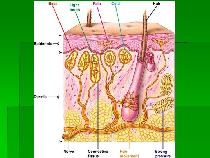
Touch § Skin contains receptors for touch & temperature § These are found in different concentrations in skin at various locations around the body. § E. g. very few in the heel of the foot compared to the elbow which has several (this is why parents use their elbow to test the temp of a babies bath.
Taste § Receptors are located in taste buds. Which are found on the top and sides of the tongue & in some parts of the throat. § 4 basic taste receptors § Sweet, bitter, sour & salt. (See diagram) § Flavour of food is combination of taste, smell, texture & temp, hence when we have a cold food looses its taste.
Taste
Smell § The nasal cavity has 20 million neurons to detect smell (olfactory neurons) § These respond to over 50 different chemicals in a gaseous state, which combine to produce 10, 000 different smells. § These receptors also adjust to a smell very quickly & stop responding. Hence a smell disappears after awhile.
Sight
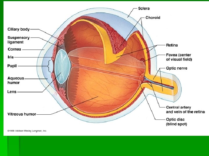
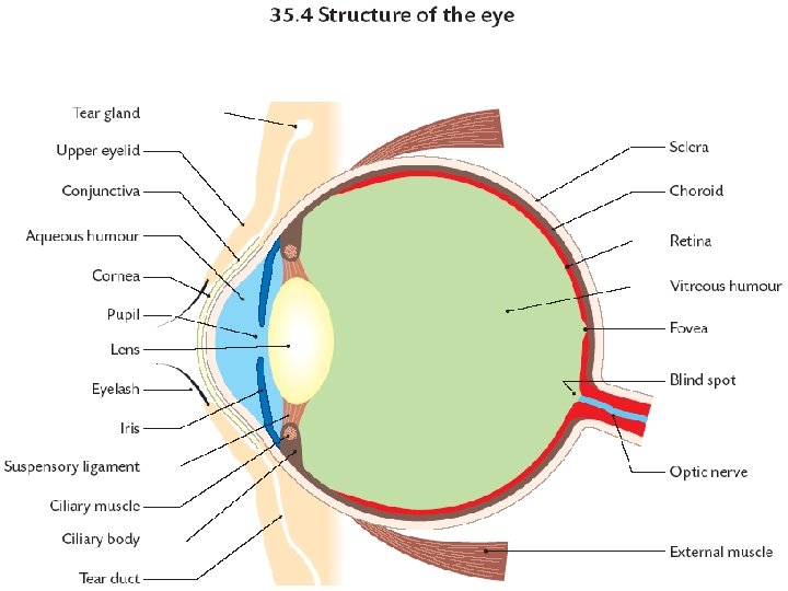
Part of Eye Function Conjuctiva Sclera Cornea Thin memb, protests sclera (inflammation=Conjunctivitis) White of eye, lets no light in, is tough & holds shape of eye Clear part of eye, allows light into eye & bends toward retina Choroid Layer which provides blood to eye & Melanin to absorb light Retina Contains light receptors (cones&rods), Pigment = rhodopsin Fovea Only contains cones, region of sharpest vision Blind Spot No rods/cones located here, nerve fibres leave eye here
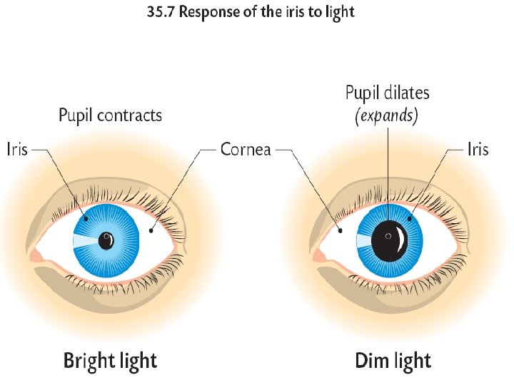
Part of Eye Function Optic Nerve Lens 126 million axons carry impulses from rods & cones to brain Changes shape to focus light on retina Ciliary Muscle Causes shape of lens to change Iris Coloured part of eye, Controls amount of light entering eye Pupil Opening in iris, lets light into eye, large opening =dilated Aqueous Salt solution which holds the shape of front of Humour the eye Vitreous Viscous fluid, supports eye by exerting pressure Humour outwards External muscle Eye is moved by 6 external muscles
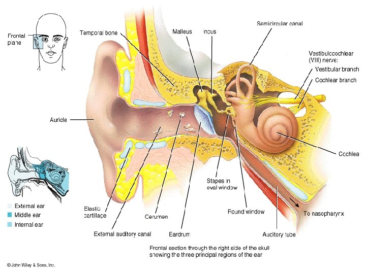
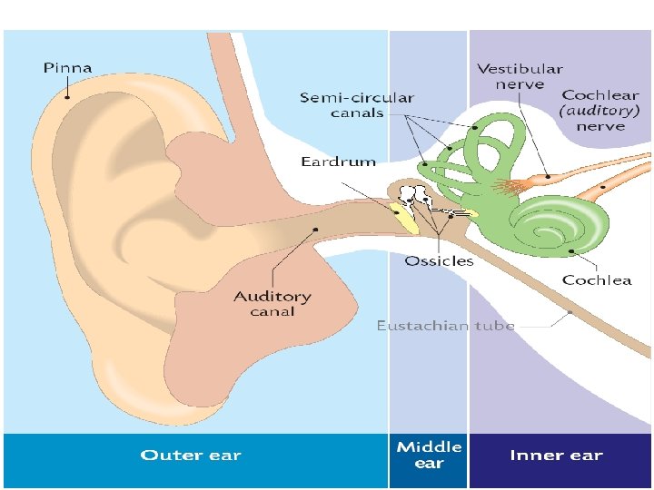
Hearing Function of the Ear § Hearing & balance § 3 sections of ear (outer, middle & inner ear) § Outer & middle ear filled with air § Inner ear filled with fluid called lymph.
The Ear & Hearing § Sound caused by vibrations in the air. § Vibrations collected by outer ear, passed through the middle ear (vibrations amplified) and transferred to fluid in the Cochlea on inner ear. § Cochlea contains receptors which are stimulated by pressure waves in lymph. These receptors cause electrical impulses to be sent to the brain which interprets them as sound.
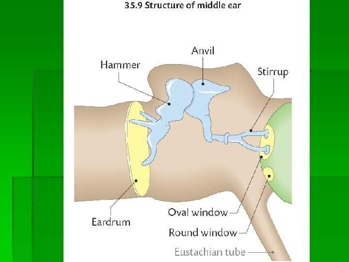
Hearing Part of Ear Function Pinna Collects & channels vibrations into Auditory canal Carries vibrations to eardrum, wax secreted outside eardrum to trap dust & protect the ear Eardrum (tympanic membrane) vibrates due to air vibrations Ossicles 3 tiny bones in middle ear that vibrate & amplify vibrations (hammer, anvil & stirrup) Eustachian tube Runs from middle ear to throat, It equalises pressure on either side of eardrum (pops when we yawn) Cochlea Spiral tube (resembles snails shell) converts pressure waves caused by vibrations into electrical impulses which travel to brain
Cochlea § Vibrations arrive at cochlea from stirrup which is attached to a membrane on the cochlea called the oval window. § Vibrations pass through OW & form pressure waves in lymph in cochlea § Pressure waves stimulate receptors which form a structure called the organ of Corti § Receptors send electrical impulses to brain Impulsestravel along auditory/cochlear nerve. § Round window allows pressure waves to dissipate out of cochlea into air of middle ear.
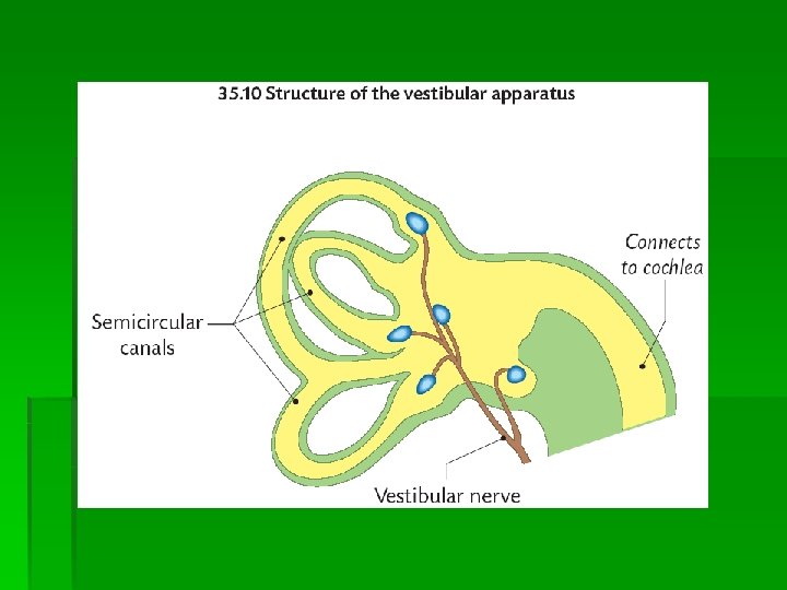
The Ear & Balance § Balance largely detected in Vestibular Apparatus in inner ear. § VA Consists of 3 semicircular canals § Person who damages VA loses sense of balance § VA is filled with Lymph § Receptors in VA detect whether head is vertical or not & other detect movement. § Receptors send impulses to cerebellum of brain through vestibular nerve.
Corrective measures for hearing disorder Disorder § Glue ear = surplus sticky fluid in middle ear which blocks the Eustachian tube & prevents movement of eardrum & small bones in middle ear Correction § Mild cases= nose drops to unblock ET § Severe cases=small tubes called grommets inserted which allow air into the middle ear & forces fluid down ET. Grommets eventually fall out themselves.