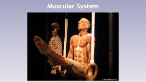
The Muscular System myo muscle Muscular System There


- Slides: 11

The Muscular System “myo = muscle”

Muscular System • • There are over 600 skeletal muscles They make up 40 -50% of our body weight Muscles play a crucial role in communication No other animal has as complex facial muscles as humans The smallest muscle in the body is the stapedius and controls the stapes (stirrup bone in the ear) The largest muscle in the body? ? ? Gluteus maximus!

Skeletal Muscle Cells • Muscle cells=muscle fibers • Layers of connective tissue group muscles fibers together and give them strength • The epimysium, perimysium, and endomysium, are continuous with each other. • Perimysium holds muscle cells together to form a fascicle. • Fascia is tough connective tissue that covers muscle organ including the epimysium

Levels of Organization of a Muscle Fascicles Muscle Fiber(cell) Myofibril Thick/Thin filaments

Shape, Size and Arrangement • Skeletal muscles are considered organs • Their arrangement and shape are suited to their function Parallel Convergent Pennate Bipennate Sphincter • They form small fibers, flat sheets or bulky masses • Long muscles have greater range of motion than short ones • Long and short fibers can contract with equal force

Origin and Insertion Points • • During contraction one bones usually stays stationary Skeletal muscles can only contract Origin is point of attachment that doesn’t move Insertion is point that does move with contraction

Muscle Actions Agonist – Also called the prime mover – Contracts to produce movement Antagonist – Performs opposing action of agonist – Relaxes during contraction Synergist – Contract at same time of agonist – Makes prime movers action more efficient Fixator muscles – – Stabilize joints Help maintain posture during movement

Lever Systems First class levers – Work like seesaws Second class levers – Work like wheelbarrow Third class levers – Flexing of forearm at elbow – Most common type in body

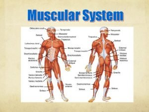
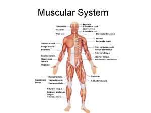
Major Muscles of the Body

How Muscles are Named Location – Brachialis(arm) & gluteus(buttocks) Action – Adductor brevis Direction of fibers – Rectus abdominis Shape – Deltoid (triangular) Number of heads/divisions – Biceps, triceps, quadriceps Origin and Insertion – Sternocleidomastoid (sternum, clavicle, mastoid) Relative Size of muscle – Gluteus maximus, medius and minimus

Quick Review • • • Connective tissue that covers individual muscle fibers? – Endomysium Connective tissue that surrounds and connects fascicles? – Perimysium Covers the muscle as a whole? – Fascia Which term includes all the rest- thin filament, muscle fiber, myofibril, fascicle? – fascicle This connects muscle to bone? – Tendon Name three fiber arrangement? – Parallel, convergent, pennate, bipennate, sphincter Point of attachment that does not move with contraction? – Origin Does move with attachment? – Insertion Muscle that directly performs a movement? – Agonist Muscles the help maintain balance? – Fixator muscles What is the most common type of lever system found in the body? – 3 rd class Name two ways muscles are named and give examples. – Points of insertions-biceps, Shape deltoid, direction of fibers-rectus abdominis, functions-adductor group, location- medialus, lateralis, intermedius

