Rodrigo Fernandes da Silva Adriana Yoshida Daniela Maira


- Slides: 3

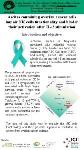
Rodrigo Fernandes da Silva; Adriana Yoshida; Daniela Maira Cardozo; Rodrigo Menezes Jales; Sophie Derchain; Fernando Guimarães. University of Campinas – Unicamp. Email: fernando@caism. unicamp. br Ascites containing ovarian cancer cells impair NK cells functionality and hinder their activation after IL-2 stimulation Introduction and objective Peritoneal ascites is frequently associated with epithelial ovarian cancer (EOC). Ascites can have free malignant cells (ASC-CA) or not (ASC). Additionally, ascites also contains soluble factors and cells from immune system, making it consistent with tumor microenvironment. CO M PE TE NC E EOC MICROENVIRONMENT High 5 -year survival rate Lymphocytes (TIL and TAL) CD 3+ CD 8+ (>5 cells/200 x field) NK Memory T-cells T-reg N IO SS RE PP SU The presence of lymphocytes in EOC has been correlated with patient outcome. CD 3+ and CD 8+ T lymphocytes are associated with high 5 -year survival, while T-regs with shortened survival and immunosuppression. Cytokines IL-10 and TGF-β, growth factors (VEGF), and more recently adenosine have also been accounted for the immunosuppression. IL-10 TGF-β ADENOSINE VEGF EOC cells Low 5 -year survival rate The goal of this study was to evaluate the NK cells functionality and their possible suppressive mediators in ascites from ovarian cancer patients. Hosted by

Rodrigo Fernandes da Silva; Adriana Yoshida; Daniela Maira Cardozo; Rodrigo Menezes Jales; Sophie Derchain; Fernando Guimarães. University of Campinas – Unicamp. Email: fernando@caism. unicamp. br Results Figure 1. Lymphocytes subtypes in ascites of patients with epithelial ovarian cancer Although T lymphocytes (CD 8 and CD 4) did not change significantly between ascites with (ASCCA) and without (ASC) malignant cells, T-regs increased in ASC-CA. Figure 2. NK activation against K 562 malignant cells and response to IL-2 Mononuclear cells (AMC) were obtained from ascites with (ASC-CA) and without (ASC) epithelial ovarian cancer cells. Resting and IL-2 stimulated AMC were challenged by co-incubation with K 562 cell lineage. The activation of NK cells in AMC was revelled by the expression of CD 107 a molecule on CD 3 -CD 56+ lymphocytes. Treatment with IL-2 had no significant effect on the activation of NK cells in ASC-CA. Basal NK cell activation in ASC was significantly higher than all other groups and remained higher after IL-2 stimulation, as if these NK cells were pre-stimulated in ASC. Differently from ASC-CA, ASC correlates to an immune competent environment. Hosted by

Rodrigo Fernandes da Silva; Adriana Yoshida; Daniela Maira Cardozo; Rodrigo Menezes Jales; Sophie Derchain; Fernando Guimarães. University of Campinas – Unicamp. Email: fernando@caism. unicamp. br Results Figure 3. Cytokine profile in ascites of patients with epithelial ovarian cancer Ascites without malignant (ASC) cells had significantly higher IL-2, which is consonant with the observation that NK cells from ASC had a better functional performance (Figure 2), as if they were pre-stimulated. Ascites with EOC cells (ASC-CA) had high levels of inflammatory cytokines TNF and IFNg. Figure 4. Comparison of the impact of T-reg and malignant EOC cells on NK function There was an inverse correlation between the amount of T-regs in ascites and the frequencies of activated NK cells. This correlation was weak, indicating that T-regs might exert a mild modulation effect on NK cells. The presence of EOC cells (black dots) was the major suppressor of NK activation in this setting. Discussion and conclusion Ascites with EOC cells had high levels of inflammatory cytokines together with the presence of T-regs and malignant cells. This is consistent with a favorable environment for the production of adenosine. In contrast to ascites without EOC cells, ascites containing malignant cells creates an immunosuppressive environment. Financial Support: FAPESP 2014/07401 -3 and 2014/17444 -1. Hosted by


