Pteris sp Occurrence and Distribution This is a

Pteris sp. - Occurrence and Distribution: • This is a cosmopolitan fern being distributed in almost all geographical regions. Pteris however, prefers tropical and sub tropical climates. Plants usually grow in well drained places or in the crevices of rocks. They are very common along the slopes of hills and can be seen even at 1200 metres above sea level. • There about 250 280 species reported for the genus. Some of the common Indian species are P. quadriaurita, P. critica, P. vittata, P. pellucida, P. wallichiana, P. stenophylla, P. biaurita, etc. Tanaka and Chin ming (1982) have isolated Pterosin and Astragalin from Pteridium acquilinum sub spp Wightianum.

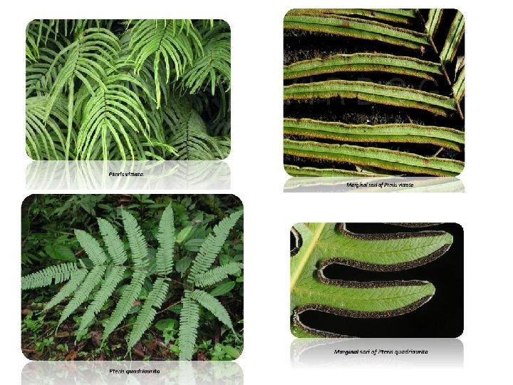
Pteris : Structure of Sporophyte: Morphology of the Plant: • The plant body has a rhizomatous stem that produces roots and leaves. The rhizome may be creeping (P. grandiflora) or compact and erect as in P. cretica and P. longifolia. The rhizome may or may not show branching and is covered with scales. Roots arise at the base of the leaf or all over the rhizome. The growing point of the rhizome is covered with ramenta. • The leaves arise from the upper surface of the rhizome and have a long rachis. They are unipinnately compound, decompound or multi pinnately compound. The dissections of the pinnae are not as deep as in Pteridium. • Venation is of open dichotomous type. The pinnae are small near the base, large towards the middle and once again small towards the apex as in P. vittata. The leaves show circinate vernation that is typical to true ferns. • Very often pinnae are Coriaceous. The leaves bear reproductive structures sorus along the ventral margin of pinnae. The sorus is continuous along the margin but avoiding the apices of the segments and usually in the sinuses between them.

Internal Structure of Sporophyte Rhizome: • The rhizome in a transection exhibits quite a good amount of diversity particularly in the stelar organisation. It is solenostelic (P. grandiflora, P. vittata), or dictyostelic. In P. biaurita the rhizome shows a mixed protostele in the lower region. It becomes siphonostelic a little up, and near the apex it shows a polycyclic dictyostelic organisation. • The epidermis is single layered and it encloses a broad cortex. There is a hypodermal sclerenchyma. The bulk of the cortex however, is made up of parenchyma. The stele consists of a number of meristeles. Usually there are two rings of meristeles. The inner ring consists of 2 or 3 large meristeles whereas the outer ring consists of a number of small meristeles. • Each meristele has a band or plate like xylem mass. Sometimes it is angular. One or two protoxylem groups are embedded in the metaxylem (mesarch). Surrounding the xylem is the phloem. Each meristele may have its own endodermis (This however should not be mistaken for the polysteles of Sehginella). Here the breaking of the vascular strand is due to the leaf gaps.

Internal Structure of Sporophyte Leaf: • The leaves are borne on the upper surface of the rhizome. When young the leaves are spirally coiled and show circinate vernation that is typical of true ferns. The leaves are unipinnately or multipinnately compound or decompound with a long rachis. • The pinnae are small near the base as well as towards the apex, while they are large towards the middle. The pinnae are very often coriaceous. All leaves are fertile, bearing sori along the ventral margin of pinnae, except the apices of the segments. • The rachis is traversed by a single C/U/ V shaped leaf trace. The lamina is bifacial, hypostomatic. Mesophyll cells may or may not be differentiated. A concentric vascular bundle with distinct bundle sheath is present in the midrib. • The rachis is traversed by a single leaf trace which is variable in shape. ‘C’ shaped leaf traces are found in P. vittata. In P. biaurita the leaf trace is ‘U’ shaped while entering the leaf base, but further up it becomes ‘V’ shaped. • The xylem strand appears hooked. As usual xylem is surrounded by phloem, pericycle and endodermis. The cortical region has an inner parenchymatous zone and an outer sclerotic zone. Epidermis is single layered with a deposition the cuticle over it. Ramenta arise from some of the epidermal cells. • The lamina appears bifacial. It may or may not have a differentiated mesophyll. Leaves are generally hypostomatic. The midrib has a concentric vascular bundle (P. vittata) with a distinct endodermis (bundle sheath).
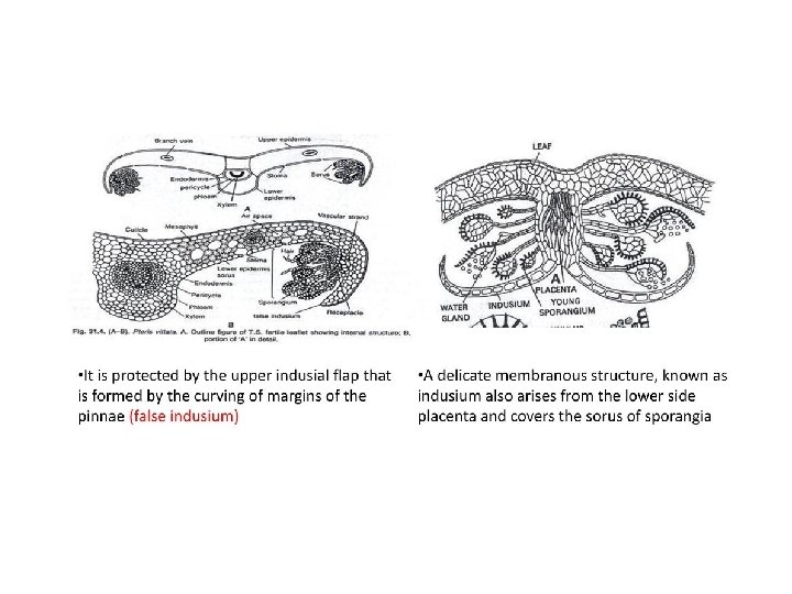

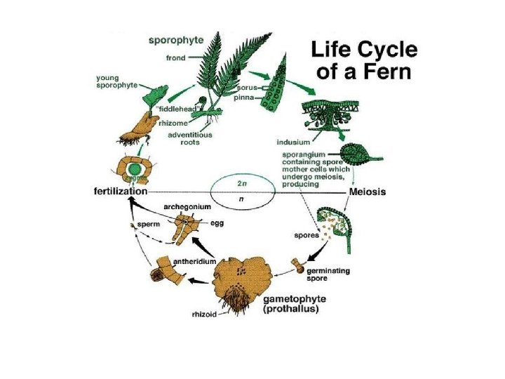
Reproduction in Pteris reproduces by means of spores. Spore-Producing Organ: • Pteris is a homosporous fern. The sorus of Pteris is called coenosorus. Coenosori are marginal, borne continuously on a vascular commissure connected with vein ends. Thus the sporangia of Pteris form a continuous linear sorus along the margin, hence the indivi duality of sori is lost. • The coenosori are protec ted by the reflexed margin (false indusium) of the pinnae. Sori are of mixed type intermingled with many sterile hairs in between the sporangia.

Structure of a Mature Sporangium • • A mature sporangium has a long stalk that terminates in a capsule The jacket of the capsule is single layered, but with three different types of cells (1)A thick walled vertical annulus incompletely overarches the sporan gium, (2) A thin walled radially arranged stomium, and (3) Large parenchymatous cells with undulated walls. The capsule contains many spores. All spores are structurally and functionally alike; hence Pteris is a homosporous pteridophyte. Spores are triangular in shape with trilete aper ture, bounded by two walls. The outer wall, exine, is variously ornamented. The sporangium dehisces transversely along the stomium due to the shrinkage of annular cells. The spores are dispersed through air to a moderate distance.

Structure of Sporangium

Development of Sporangium • The development of sporangium in Pteris is of leptosporangiate type. A single superficial cell of the receptacle functions as the sporangial initial which divides transversely to produce an upper cell and a lower cell. • The lower cell does not take part in sporangium development, while the upper cell, by intersecting oblique walls, gets differentiated into an apical cell with three cutting faces. The apical cell cuts off two segments along each of its three cutting faces. • The apical cell divides periclinally to form an outer jacket initial and an inner tetrahedral archesporial cell. The jacket initial divides, anticlinally to form a single layered jacket of the sporangium. The archesporial cell further divides periclinally to form an outer tapetal initial and an inner primary sporogenous cell. • The tapetal initial by one periclinal and several anticlinal divisions forms two layered tapetum. The primary sporogenous cell divides to form 12 spore mother cells. The spore mother cells divide meiotically to produce haploid spores, while the tapetal cells disorganise and provide nutrition to the spores.

Dehiscence of Sporangium

Structure of Gametophyte • The spores germinate after falling on a sui table substratum. Initially the spore wall (exine) ruptures and the inner contents come out in the form of a germ tube and subsequently by a transverse division in the germ tube forms the first rhizoid and the first prothallial cell. The prothallial cell divides to form a small filament having an apical terminal cell with two cutting faces. • The apical cell further divides and a spathulate prothallus is formed first. Finally a mature prothallus is formed which becomes cordate, dorsiventrally flattened, aerial and photosynthetic. • The prothallus is made up of parenchymatous cells which are single celled thick towards the margin and many celled thick towards the centre. The grow ing point are located in the apical notch. Rhizoids are formed over the ventral surface. • The prothallus is monoecious, protandrous. Antheridia appear first and are confined to the basal central or lateral regions among the rhizoids. Archegonia develop near the apical notch. .

Development and Structure of the Antheridium • • One of the superficial cells on the prothallus towards the ventral surface projects out a little and functions as an antheridial initial. This undergoes a transverse division to form two superposed cells. Due to the increase of turgor pressure in the upper cell, the cross wall bulges down and ultimately touches the wall of the lower cell. The upper cell now divides to form a dome cell and the primary androgonial cell. At this stage the young antheridium consists of a dome cell, a primary androgonial cell and a ring cell (lower cell looking like a ring). The dome cell divides transversely to form a cap cell and the second ring cell. The cap cell may are may not divide to form two cover cells. Meanwhile the primary androgonial cell divides to form 20 25 androcytes. A mature antheridium has two ring cells, one or two cover cells enclosing a mass of antherozoids which are coiled and multi ciliate. At maturity the antheridium absorbs water and swells, the cover cell opens out releasing the an therozoids.

Development and Structure of the Archegonium: • The archegonium is also initiated in the derivates of apical meristem. One of the superficial cells functions as archegonial initial which, on periclinal division, forms an outer primary cover cell and an inner cell. The primary cover cell, by two vertical divisions at right angles to each other, forms quadrant of neck initials. • Further anticlinal divisions of neck initials form the neck of the archegonium. The neck barely protrudes out of the thallus. The inner cell, on the other hand, divides periclinally (transverse) to form a basal cell and a central cell. • The central cell divides transversely into a primary canal cell and a primary venter cell. The primary canal cell directly functions as neck canal cell. The primary venter cell, however, divides transversely and forms a ventral canal cell and a large egg. • At maturity, the ventral canal cell, the neck canal cell and the neck cells at the top are well disorganised, they thus form an open passage for the antherozoids to come towards the egg • A mature archegonium of Pteris consists of a 5 6 celled projecting curved neck, a neck canal cell, a ventral canal cell and an egg. .

Development of New Sporophyte Fertilisation: The antheridium at maturity absorbs water and swells. Due to the increase in pressure with in the antheridium the cover cells split apart releasing the antherozoids in a thin film of water present on the surface of the prothallus. • At the same time the ventral canal cell, the neck canal cell and the neck cells at the top disintegrate forming an open passage for the antherozoids to come towards the egg and, eventually, one of the antherozoids fuses with the egg to form the zygote • New Sporophyte (Embryo): • Like other leptosporangiate ferns, in Pteris the first division of the zygote is vertical, followed by a second transverse division resulting in the formation of a quadrant. Further a 32 celled embryo is formed due to further divisions of the quadrant. • The differentiation of embryo begins at this 32 celled stage. No suspensor is formed; the hypobasal cells form stem apex and foot, while epibasal cells form cotyledon and root. With the development of embryo, the venter of the archegonium forms a protective layer, called catyptra, around the embryo. • In the young embryo the root and cotyledon grow more rapidly than the shoot. The root pierces the prothallus and establishes the sporeling in the soil. Later, the first leaf develops.

Life Cycle of Pteris
- Slides: 17