Microscopy Notes Scales of measurement Nanometer nm 1

: 1 billionth of a meter ● Micrometer (um):")
 • Organelles (up to 10 um)")


 • Inexpensive and easy to use • Used")
 • Can magnify up to 250, 000 times. This")


- Slides: 16
Microscopy Notes
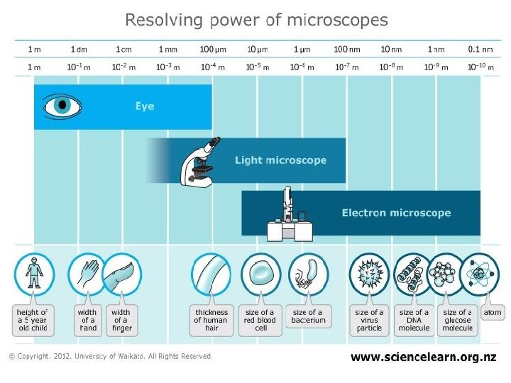
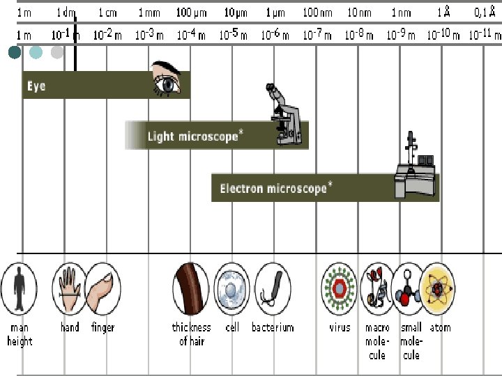
Scales of measurement ● Nanometer (nm): 1 billionth of a meter ● Micrometer (um): 1 millionth of a meter ● Millimeter (mm): 1 thousandth of a meter ● Centimeter (cm): 1 hundredth of a meter
Relative sizes • Cells (up to 100 um) • Organelles (up to 10 um) • Bacteria (1 um) • Viruses (100 nm) • • Cell membrane thickness (10 nm) Molecules (1 nm) (large to small)
Microscopes are instruments that can magnify and resolve objects • Magnification ● How much larger the object appears compared to its real size.
What is different about the photo quality?
Microscopes are instruments that can magnify and resolve objects • Resolution ● The ability to form separate images of objects that are very close together. ● Resolving power is stated as the minimum distance two points can be separated and still be distinguished as two separate points. ● The lower the resolving power, the better the resolution
Light microscopes (use lenses and light) • Inexpensive and easy to use • Used to study stained or living cells in color • Objects can be magnified up to 2000 X Ours at school only magnify 1000 X • Can resolve objects 200 nm apart (500 times better than the human eye)
Electron microscopes (use electron beams) • Can magnify up to 250, 000 times. This is 125 times the magnifying power of light microscopes. • Can resolve objects that are 0. 2 nm apart. This is 1000 times the resolving power of light microscopes. • Requires cells to be killed and chemically treated before viewing. • No color can be seen
Cell Structure Light Microscope Electron Microscope Membrane YES – but not in much detail YES Ribosome NO YES (if stained) YES Golgi apparatus NO YES Endoplasmic Reticulum NO YES Mitochondria Chloroplast Cytoskeleton YES TRY YES ON YOUR NO OWN ISFIRST!! IT YES Nucleus VISIBLE? YES YES OR YES NO? YES Cell wall YES Flagella YES Lysosome YES – but hard to distinguish Vacuole YES – but hard to distinguish YES
Cell Structure Light Microscope Electron Microscope Membrane YES – but not in much detail YES Ribosome NO YES (if stained) YES Golgi apparatus NO YES Endoplasmic Reticulum NO YES YES YES Lysosome YES – but hard to distinguish YES Vacuole YES – but hard to distinguish YES Nucleus YES Cell wall YES Mitochondria Chloroplast Cytoskeleton Flagella
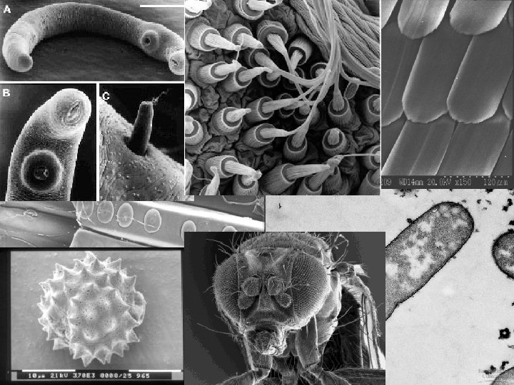
Micrographs are pictures taken through microscopes
Developments in Scientific Research Follow Improvements in Apparatus • Use the next slide and your own research to outline how advances in microscopy led to a greater understanding of cell structure. • Make sure to touch on what limitations the light microscope has and why it has them, as well as how the electron microscope is able to reveal a greater deal of resolution.
Use these 1. 2. The Article “Scrutinizing the Microcosm” The Article “The Invention of the Electron Microscope”