Mammalian Anatomy Human Anatomy Identify the following parts


- Slides: 9

Mammalian Anatomy

Human Anatomy Identify the following parts: Digestive (slide 3) • • Esophagus Stomach Small intestine Large intestine Rectum Pancreas (accessory organ) Liver (accessory organ) Gall Bladder(accessory organ) Respiratory(Slide 4) • • • Trachea Lung Bronchus Bronchioles Alveoli Diaphragm Heart (Slide 6) • Right and Left Atrium • Right and left Ventricle • Tricuspid and Bicuspid valve Urinary (Slide 7) • Kidney • Ureter • Urinary Bladder Histology • Villi of small intestine (Slide 8) • Red Blood cell (Slide 9) • White Blood Cells (Slie 9)

**Figure 24. 2 The digestive system, or the gastrointestinal tract, includes all of the organs associated with the digestion of food. Digestive System: Mouth: for ingestion, chewing and saliva that contains enzymes and mucous Pharynx: muscular organ that forces food down the digestive tract Stomach: mixes food with acid and enzymes Small intestine: absorbs food Large intestine: reabsorbs water and houses flora Liver: produces bile to emulsify lipids Pancreas: releases enzymes and bicarbonate into the small intestine

**Figure 22. 3 The structures of the lower respiratory tract are identified in this illustration. (credit: modification of work by National Cancer Institute) The respiratory system: The Larynx (voice box) leads to the Trachea that bifurcates into two Bronchi that bifurcate into smaller Bronchioles that end in Alveolar Sacs for increased surface area for gas exchange.

**Figure 22. 4 This micrograph shows the structure of the mucous membrane of the respiratory tract. (credit: modification of micrograph provided by the Regents of University of Michigan Medical School © 2012) Trachea: ciliated, pseudostratified, columnar epithelium

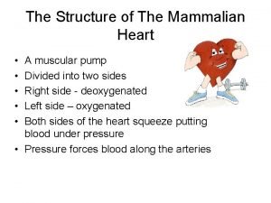
**Figure 25. 2 The major components of the human circulatory system include the heart, arteries, veins, and capillaries. This network delivers blood to the body’s organs and tissues. (credit top left: modification of work by Mariana Ruiz Villareal; credit bottom right: modification of work by Bruce Blaus) Heart: Superior & Inferior Vena Cava drain blood into Right Atrium that pumps blood to the Right Ventricle that pumps blood to the Lungs to become oxygenated Left Atrium pumps blood to the Left Ventricle that pumps blood to the Aortic Artery that carries blood to the body

**Figure 23. 2 These structures of the human urinary system are present in both males and females. Excretory System: Renal Artery brings blood to the kidneys, Renal Vein brings blood to the veins Kidneys filter blood, Ureters carry blood to the bladder Bladder stores urine, Urethra carries urine outside the body

**Figure 24. 5 (a) The structure of the wall of the small intestine allows for the majority of nutrient absorption in the body. (b) Villi are folds in the surface of the small intestine. Microvilli are cytoplasmic extensions on individual cells that increase the surface area for absorption. (c) A light Small intestine: micrograph shows the shape Villi increase surface area of the villi. (d) An electron micrograph shows the shape of the microvilli. (credit b, c, d: Modification of micrographs provided by the Regents of University of Michigan Medical School © 2012)

Figure 42. 1 In this compound light micrograph purple-stained neutrophil (upper left) and eosinophil (lower right) are white blood cells that float among red blood cells in this blood smear. Neutrophils provide an early, rapid, and nonspecific defense against invading pathogens. Eosinophils play a variety of roles in the immune response. Red blood cells are about 7– 8 µm in diameter, and a neutrophil is about 10– 12µm. (credit: modification of work by Dr. David Csaba) White blood cells with multi-lobed nuclei Red Blood Cells

