Life is Cellular Chapter 7 1 The Discovery

- Slides: 19
Life is Cellular Chapter 7. 1
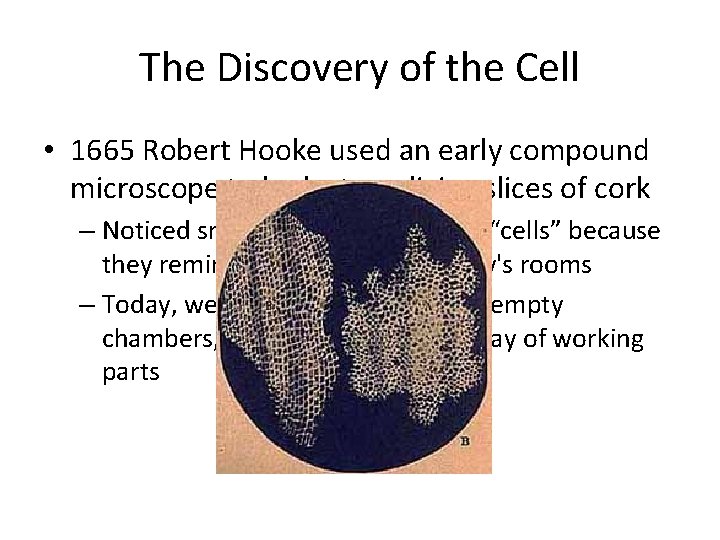
The Discovery of the Cell • 1665 Robert Hooke used an early compound microscope to look at nonliving slices of cork – Noticed small chambers he called “cells” because they reminded him of a monastery's rooms – Today, we know that cells are not empty chambers, they contain a huge array of working parts
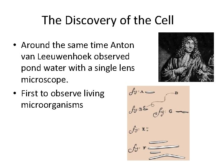
The Discovery of the Cell • Around the same time Anton van Leeuwenhoek observed pond water with a single lens microscope. • First to observe living microorganisms
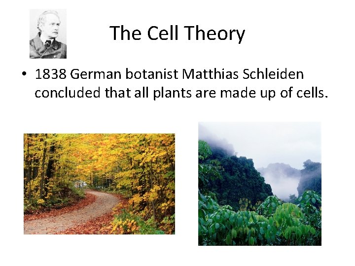
The Cell Theory • 1838 German botanist Matthias Schleiden concluded that all plants are made up of cells.
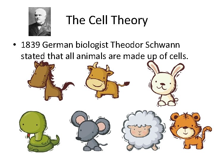
The Cell Theory • 1839 German biologist Theodor Schwann stated that all animals are made up of cells.
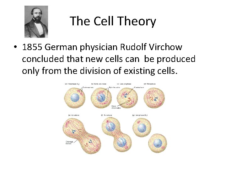
The Cell Theory • 1855 German physician Rudolf Virchow concluded that new cells can be produced only from the division of existing cells.
The Cell Theory states: • All living things are made up of cells. • Cells are the basic units of structure and function in living things. • New cells are produced from existing cells. The cell theory
Exploring the Cell • Most microscopes use lenses to magnify the image of an object by focusing light or electrons.
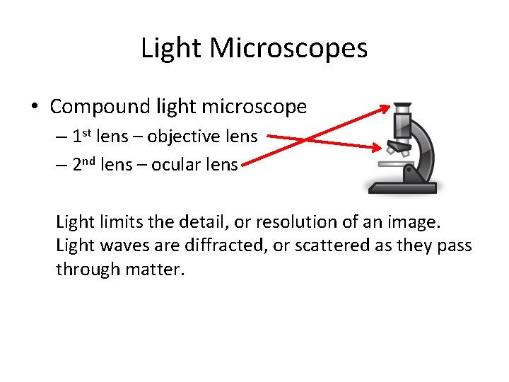
Light Microscopes • Compound light microscope – 1 st lens – objective lens – 2 nd lens – ocular lens Light limits the detail, or resolution of an image. Light waves are diffracted, or scattered as they pass through matter.
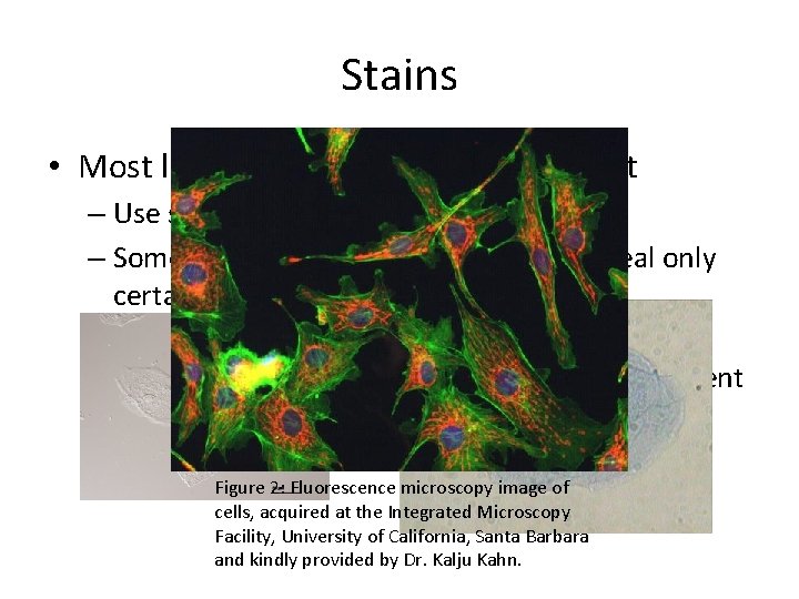
Stains • Most living cells are nearly transparent – Use stains or dyes to solve this problem – Some stains are so specific that they reveal only certain compounds or structures – Fluorescent dyes can be attached to specific molecules and be made visible using a fluorescent microscope Figure 2: Fluorescence microscopy image of cells, acquired at the Integrated Microscopy Facility, University of California, Santa Barbara and kindly provided by Dr. Kalju Kahn.
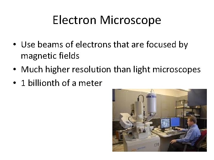
Electron Microscope • Use beams of electrons that are focused by magnetic fields • Much higher resolution than light microscopes • 1 billionth of a meter
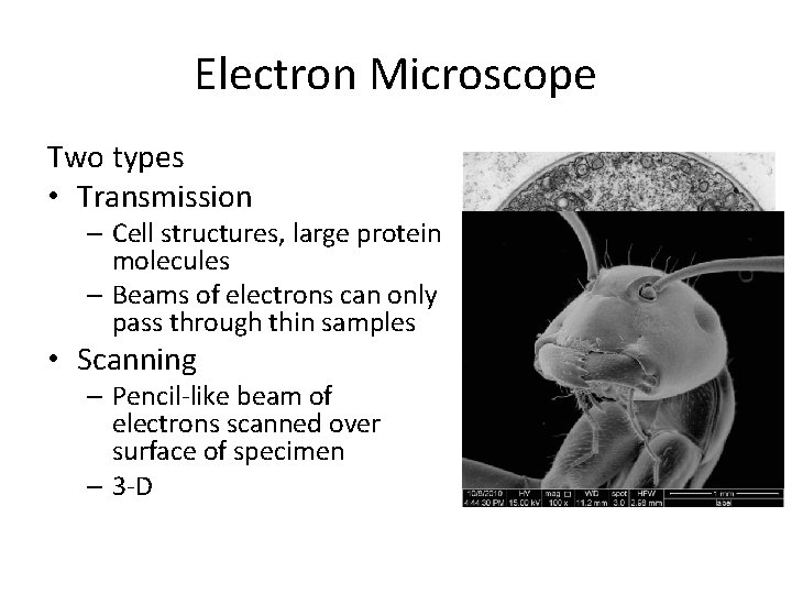
Electron Microscope Two types • Transmission – Cell structures, large protein molecules – Beams of electrons can only pass through thin samples • Scanning – Pencil-like beam of electrons scanned over surface of specimen – 3 -D
Electron Microscopes Since electrons are easily scattered by the air, samples must be placed in a vacuum. Scientists must chemically preserve their samples…only non-living cells and tissues.
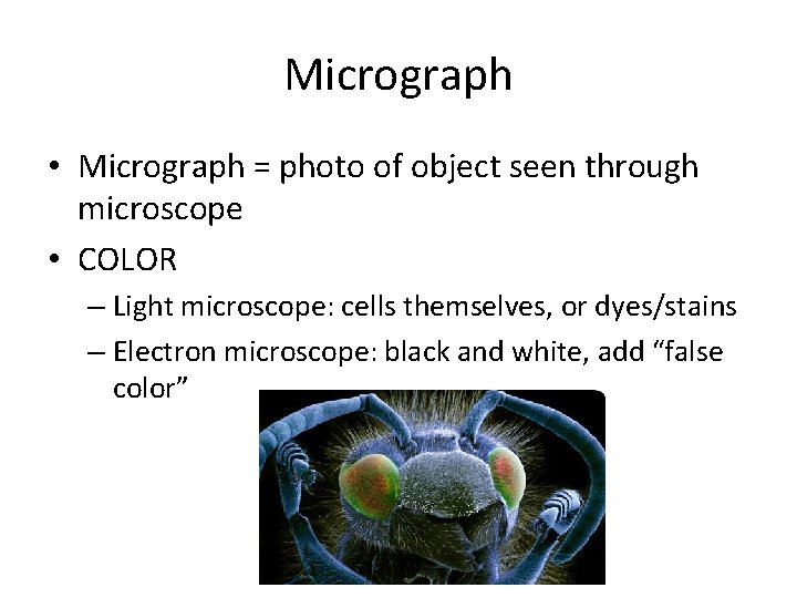
Micrograph • Micrograph = photo of object seen through microscope • COLOR – Light microscope: cells themselves, or dyes/stains – Electron microscope: black and white, add “false color”
Prokaryotes & Eukaryotes • Cells come in an amazing variety of shapes and sizes • All cells contain DNA- the molecule that carries genetic information • All cells are surrounded by a thin flexible barrier called a cell membrane
• Cells fall into two categories depending on whether they contain a nucleus. • Nucleus = a large membrane enclosed structure that contains genetic material in the form of DNA and controls many of the cell’s activities.
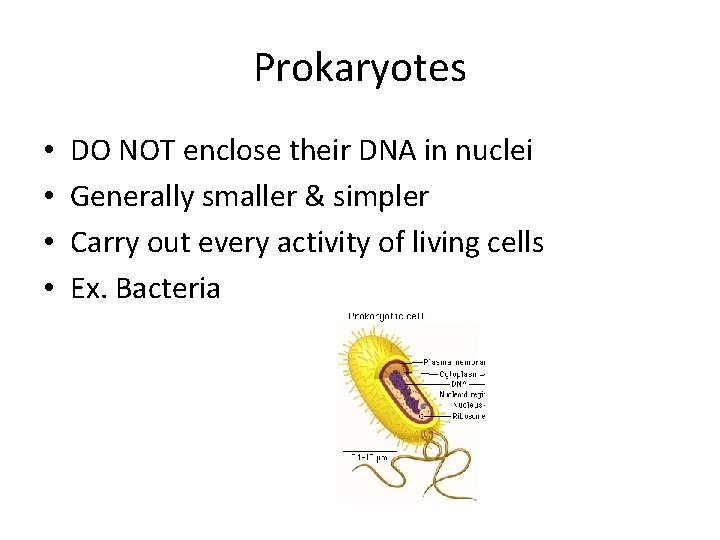
Prokaryotes • • DO NOT enclose their DNA in nuclei Generally smaller & simpler Carry out every activity of living cells Ex. Bacteria
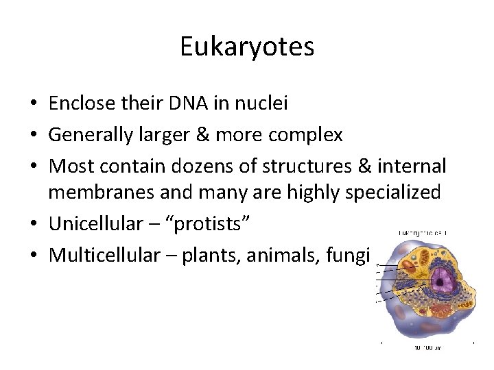
Eukaryotes • Enclose their DNA in nuclei • Generally larger & more complex • Most contain dozens of structures & internal membranes and many are highly specialized • Unicellular – “protists” • Multicellular – plants, animals, fungi
• Prokaryotes vs. Eukaryotes