Janica E Walden MD Neuroradiology University of North

- Slides: 21
Janica E. Walden, MD Neuroradiology University of North Carolina
Holoprosencephaly (HPE) �Spectrum of congenital structural forebrain anomalies defined by different degrees of frontal lobe fusion �Impaired midline cleavage of the embryonic forebrain �“Face predicts brain”: severe midline anomaly = severe HPE �Clinical severity relates to degree of hemispheric and deep gray nuclei fusion
Etiology & Pathology �Normal prosencephalic cleavage occurs at 4 -6 weeks �HPE: disruption in dorsoventral axis patterning of secondary prosencephalon, �Result of mutations affecting signaling genes (Sonic hedgehog gene) which regulate neural tube patterning. �Extreme hypoplasia of neocortex �Dorsal cyst (especially in association with non-cleaved thalami) thought to represent expansion of partially blocked posterodorsal 3 rd ventricle �Variable degree of fusion of diencephalon & basal ganglia/thalamus with incorporation into upper brainstem
Epidemiology �Occurs in 1 to 1. 4 per 10, 000 live births �More common in early embryogenesis with high spontaneous miscarriage rates �Maternal factors include alcohol use, diabetes, retinoic acid � 1% risk to infants of diabetic mothers (200 -fold increased risk than that of general population) �Male: female ratio = 1. 4: 1
Facial Anomalies �Severe facial anomalies correlate with severity of HPE in 80% �+/- midline clefting �premaxillary agenesis if severe �absent superior frenulum �+/- central incisor �proboscis �single nare; single nasal bone/absent inter-nasal sutures �caudal metopic suture �infants of diabetic mothers may have alobar HPE with near-normal facies
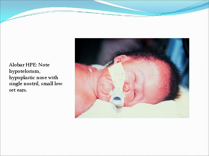
Alobar HPE: Note hypotelorism, hypoplastic nose with single nostril, small low set ears.
Clinical Features �Most severe (classic alobar HPE) features include: cyclopia, proboscis, midline facial clefting, microcephaly �Severe of pituitary/hypothalamic dysfunction (75% especially diabetes insipidus) & disturbed body temperature regulation �Correlates with degree of hypothalamic non-separation �Seizures (50%) & mental retardation �Most severe with cortical malformations �Dystonia & hypotonia �Severity correlates with degree basal ganglia non-separation
Classification �Defined by degree of frontal lobe fusion �Sylvian angle (of Barkovich) = lines drawn tangentially through Sylvian fissures �Anteriorly displaced Sylvian fissures results in increased Sylvian angle �The larger the Sylvian angle is the more severe frontal lobe hypoplasia is too � 3 types of HPE based on criteria (lobar, semilobar, and alobar), as well as a middle interhemispheric variant, septooptic dysplasia, and single central incisor
Alobar Holoprosencephaly �“Pancake” or “horseshoe” brain �Monoventricle �Large dorsal cyst �Fused diencephalon �Basal ganglia & thalami may form gray matter fusion mass �No interhemispheric fissure �No olfactory nerves
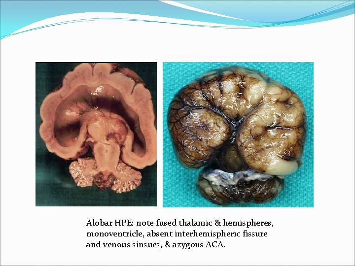
Alobar HPE: note fused thalamic & hemispheres, monoventricle, absent interhemispheric fissure and venous sinsues, & azygous ACA.
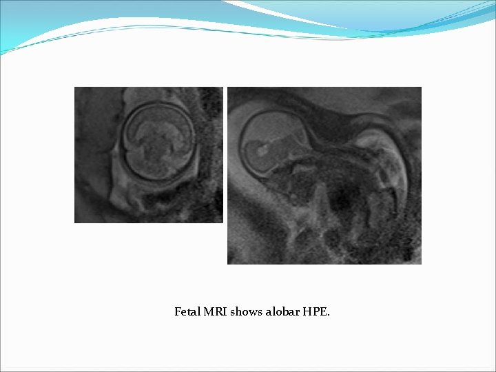
Fetal MRI shows alobar HPE.
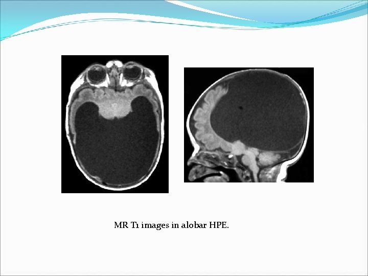
MR T 1 images in alobar HPE.
Diagnosis of HPE by Ultrasound �Diagnosis of HPE by ultrasound can be made as early as 9 weeks gestational age. �Development of forebrain can be delineated in detail with ultrasound from 7 weeks on. �Alobar HPE may be detectable as early as the end of week 7 �Non-visualization of the butterfly sign is very helpful in diagnosis
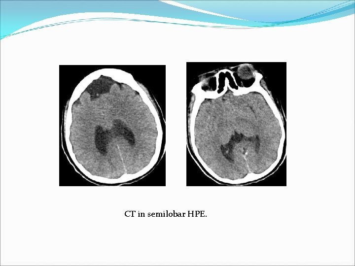
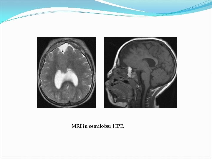
Semilobar Holoprosencephaly �Partial occipital/temporal horns �Moderate sized dorsal cyst �Fused diencephalon �Partial fusion of basal ganglia > thalami �Interhemipheric fissure present posteriorly �Absent of hypoplastic olfactory tracts and bulbs �Corpus callosum is rudimentary
CT in semilobar HPE.
MRI in semilobar HPE.
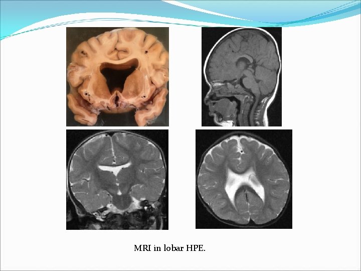
Lobar Holoprosencephaly �Formed lateral ventricles �Small or no dorsal cyst �Fused diencephalon and/or fornices �+/- partial fusion of basal ganglia > thalami �Interhemispheric fissure nearly normal �Small or normal olfactory nerves
MRI in lobar HPE.
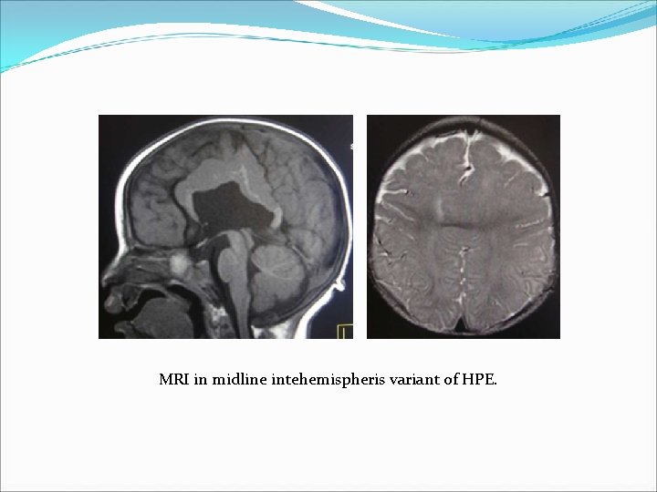
Middle Interhemispheric Variant �Sylvian fissures connect across midline over vertex (86%) �Interhemispheric fusion of posterior frontal/parietal lobes, with normal separation of anterior frontal/occipital lobes �Non-cleavage of thalami > basal ganglia �Heterotopias and cortical dysplasias common (86%) �Thought to reflect abnormal induction of embryonic roof plate �Classic HPE = abnormal induction of embryonic floor plate �May explain absence of craniofacial malformations �Spasticity, hypotonia, seizures, developmental delay
MRI in midline intehemispheris variant of HPE.
References � Sepulveda Waldo, Dezerega Victor, Be Cecilia. First-Trimester Sonographic Diagnosis of Holoprosencephaly. Journal of Ultrasound in Medicine 23: 761 -765. � Hahn Jin, Barnes Patrick. Neuroimaging Advances in Holoprosencephaly: Refining the Spectrum of the Midline Malformation. American Journal of Medical Genetics 154 C: 120 -132. � Blaas H. , Eriksson A. , Salvesen K. , et al. Brains and faces in holoprosencephaly: pre- and postnatal description in 30 cases. Ultrasound Obstet Gynecol 2002; 19: 24 -38. � Takanashi Jun-ichi, Barkovich A. James, Clegg Nancy, Delgado Mauricio. Middle Interhemispheric Bariant of Holoprosencephaly Associated with Diffuse Polymicrogyria. AJNR 2003; 24: 394 -397. � Simon Erin, Hevner Robert, Pinter Joseph, et al. The Middle Interhemispheric Variant of Holoprosencephaly. AJNR 2002; 23: 151155.