Intraoperation Arthrodesis of the DIJ joint Arthrodesis of

- Slides: 9
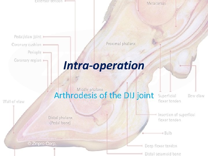
Intra-operation Arthrodesis of the DIJ joint
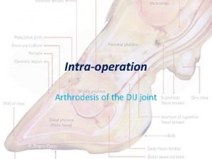
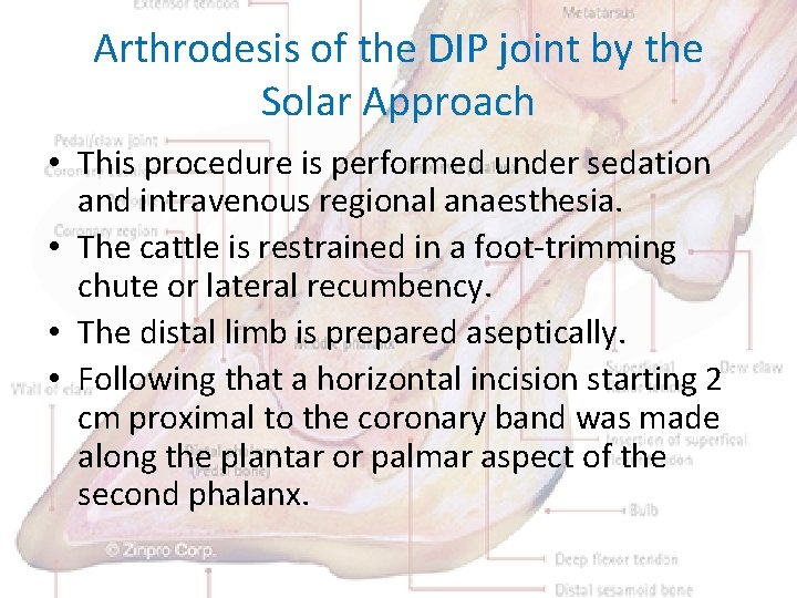
Arthrodesis of the DIP joint by the Solar Approach • This procedure is performed under sedation and intravenous regional anaesthesia. • The cattle is restrained in a foot-trimming chute or lateral recumbency. • The distal limb is prepared aseptically. • Following that a horizontal incision starting 2 cm proximal to the coronary band was made along the plantar or palmar aspect of the second phalanx.
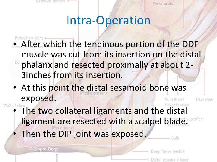
Intra-Operation • After which the tendinous portion of the DDF muscle was cut from its insertion on the distal phalanx and resected proximally at about 23 inches from its insertion. • At this point the distal sesamoid bone was exposed. • The two collateral ligaments and the distal ligament are resected with a scalpel blade. • Then the DIP joint was exposed.
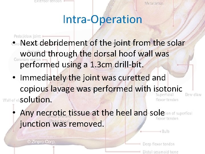
Intra-Operation • Next debridement of the joint from the solar wound through the dorsal hoof wall was performed using a 1. 3 cm drill-bit. • Immediately the joint was curetted and copious lavage was performed with isotonic solution. • Any necrotic tissue at the heel and sole junction was removed.
Intra-Operation • A wooden block was placed on the healthy digit of the affected hoof. • Then the claws are wired together with the affected digit in slight flexion. • The wound is bandaged and lavage s performed every other day. • Systematic antibiotics was recommended for 2 -3 weeks and phenylbutazone is given as needed for the first 2 weeks.
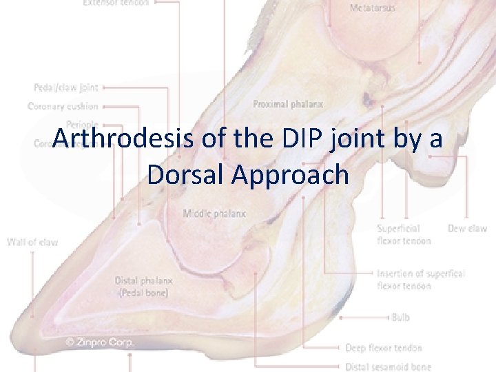
Arthrodesis of the DIP joint by a Dorsal Approach
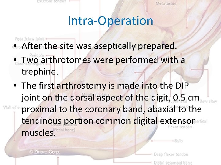
Intra-Operation • After the site was aseptically prepared. • Two arthrotomes were performed with a trephine. • The first arthrostomy is made into the DIP joint on the dorsal aspect of the digit, 0. 5 cm proximal to the coronary band, abaxial to the tendinous portion common digital extensor muscles.
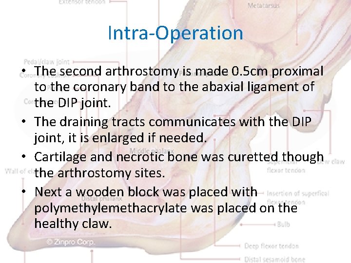
Intra-Operation • The second arthrostomy is made 0. 5 cm proximal to the coronary band to the abaxial ligament of the DIP joint. • The draining tracts communicates with the DIP joint, it is enlarged if needed. • Cartilage and necrotic bone was curetted though the arthrostomy sites. • Next a wooden block was placed with polymethylemethacrylate was placed on the healthy claw.
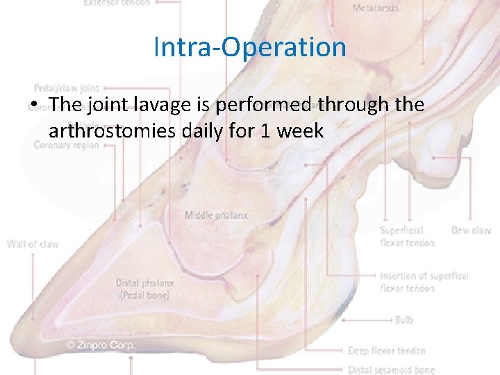
Intra-Operation • The joint lavage is performed through the arthrostomies daily for 1 week