Immunity 6 1 Defence mechanisms 6 2 Phagocytosis


- Slides: 11

Immunity 6. 1 Defence mechanisms 6. 2 Phagocytosis

Learning outcomes Students should understand the following: �Phagocytosis and the role of lysosomes and lysosomal enzymes in the subsequent destruction of ingested pathogens. �Immune system - barriers - Brainpop. swf

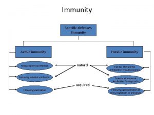
Defence mechanisms Non-specific Response is immediate and the same for all pathogens Physical barrier e. g. skin Phagocytosis Specific Response is slower and specific to each pathogen Cell-mediated response T lymphocytes Humoral response B lymphcytes

Recognising your own cells The body needs to be able to distinguish between its own cells (self) and foreign cells (non-self). In the fetus the lymphocytes (a type of white blood cell) are constantly colliding almost exclusively with the body’s own material (self). These lymphocytes are destroyed or suppresses so that the only remaining lymphocytes are those which recognise foreign (non-self) material.

Important points to remember �Specific lymphocytes are not produced in response to an infection they already exist in the body. �Given the high number of lymphocytes (10 million) their is a high probability that one of them will ‘recognise’ the pathogen. �There are only a few of each type of lymphocyte so it takes time for the body to build up the numbers of lymphocytes to destroy the pathogen, hence the time lag between infection and control.

Non-specific mechanisms Barriers to disease �Epidermis of skin �Layers of dead cells prevent invasion �Mucus membranes �Protective mucus layer secreted by goblet cells. Invaders get trapped in the mucus �Ciliated epithelia �Sweeps invaders away so they can be removed eg in the lung �Hydrochloric acid in stomach �Low p. H so the enzymes of pathogens are denatured

Problems with barriers �Some pathogens can penetrate barriers: ¬Malaria is caused by Plasmodium, which passes through the skin when a mosquito bites ¬Bubonic plaque enters the skin through flea bites ¬Influenza virus passes through lining of trachea and lungs

Phagocytosis �Phagocytosis is the process by which pathogens are taken up and destroyed by white blood cells (leucoytes). �These white blood cells are continually produced from stem cells in bone marrow � They are stored in bone marrow and released into the blood to engulf and digest foreign bodies �There are 2 types of phagocytes : Neutrophils Macrophages (monocytes)

Phagocytosis

Phagocytosis �Phagocytosis causes inflammation at the site of infection. �The swollen area contains dead bacteria and phagocytes, known as pus. �Inflammation is the result of the release of histamine, which causes dilation of the blood vessels in order to speed up the delivery of antibodies and white blood cells to the site of infection.

Learning outcomes Students should understand the following: �Phagocytosis and the role of lysosomes and lysosomal enzymes in the subsequent destruction of ingested pathogens.
 Difference between acquired immunity and innate immunity
 Phagocytosis active transport
 Seratonin
 Phagocytosis steps
 Transport mechanism
 Phagocytosis
 Smooth endoplasmic reticulum function
 Diffusion
 Cell transport
 Unfertilized ovum disappears by phagocytosis.
 Autophagy vs phagocytosis
 Tetrahymena phagocytosis lab report

