Human Physiology Nerves Homeostasis and Hormones Maintaining Homeostasis


- Slides: 29

Human Physiology Nerves, Homeostasis and Hormones

Maintaining Homeostasis �The nervous system maintains homeostasis by: • Receptor or sensor monitors the level of a variable • Coordinating centre (CNS) regulates level of the variable • Effector structures bring about the changes directed by the coordinating center, to maintain the level of the variable.

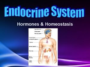
Maintaining Homeostasis �The response can be carried out by a nervous response, which is a nerve impulse (Nervous System) �The response can be carried out by the release of a hormone, acting on organs in the body (Endocrine System)

Maintaining Homeostasis �Nervous System consists of: • Central Nervous System (CNS) consisting of the brain and spinal cord • Peripheral nerves, called neurons. �Their function is to transport messages in the form of electrical impulses to specific sites. • Breathing Rate is controlled by the Nervous System • Thermoregulation is controlled by the Nervous System and Endocrine System

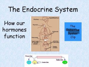
Maintaining Homeostasis �Endocrine System consists of: • endocrine glands �produce hormones to the blood (ex. Adrenal glands on the top of the kidneys. ) �do not release their product into a duct, like exocrine glands (in the digestive system). �considered ductless glands. They secrete their hormones into the blood, which transports it around the body. �Hormones act on organs, when they come in contact with target cells

Nervous System and Impulse �Nervous System • Central Nervous System �Brain and Spinal Cord �Peripheral Nervous System • Voluntary Nerves (somatic) • Autonomic Nerves (visceral)

Nervous System and Impulse �Motor Neuron • Consists of: �axon �Schwann cells, which provide a multi-layered lipid and protein coating called a myelin sheath �nodes of Ranvier. �The axon terminates at a motor end plate, or axon terminal

Motor Neuron

Nervous System and Impulse � Typical Nervous System Pathway • Starts off with a stimulus • Creates an action potential that flows from the sensory neurons to relay neurons to the brain, which interprets the stimulus • The brain sends a response through relay neurons to a motor neuron, which ends in an effector organ, like a muscle, or endocrine gland

Nerve Impulse Resting Potential (RMP) �Nerve is at rest �Maintains a more positive charge on the outside and a more negative on the inside �Nerve is said to be polarized �Charge of -70 m. V �Maintained by greater concentration of Na+ outside the cell compared to K+ and Cl- on the inside and the fact the membrane is more permeable to K+, causing it to leak out, maintaining a negative charge on the inside and positive charge on the outside

Nerve Impulse

Nerve Impulse �Action Potential Caused by a stimulus Stimulus causes depolarization to occur Neuron repolarizes All or None Principle is followed and every action potential is the same size, following the same pattern • Size of stimulus determines how many neurons are stimulated, to carry the message • •

�Progression of Action Potential • Stimulus causes the membrane sodium pores to • • • open Sodium pores allow sodium ions to flow in, reducing the charge on the inside This is called depolarization Na+ ions continue to move in by diffusion Once 40 m. V is reached, sodium pores close Neuron cannot conduct another impulse until it resets (repolarizes) Potassium channels open and K+ ions flow out, reducing positive charge on the inside of the axon

• • This is called repolarization Once axon is polarized, the potassium pores close Ion are in wrong place, so the have to be reset Done by Na+/K+ Pump (Active Transport) � Summary of Action Potential (Impulse) • The action potential is the time of depolarization (1 msec). • The refractory period is the time taken for repolarization. �Refactory period is divided into the absolute refractory state (1 msec), followed by the relative refractory state (up to 10 msec. )

Nerve Impulse – Action Potential

Myelinated vs. Non-Myelinated Axon �Myelinated nerves conduct faster than non -myelinated nerves �At nodes of Ranvier, the sodium channels are present �When impulse travels, it travels from node to node, jumping, conducting faster �Called saltatory conduction

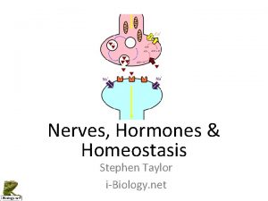
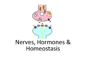
Synaptic Transmission �Like wires, there are points that join neuron to neuron, neuron to cell body, neuron to effector organ �Connection points are called synapses �Two types of synapses: • Electrical • Chemical

Synaptic Transmission �Conduction across the synapse is achieved by a neurotransmitter �Depolarization in the pre-synaptic bulb releases Ca+2, which stimulates the release of neurotransmitter �When neurotransmitter flows across the synaptic cleft, caused depolariztion of the post-synaptic bulb, to continue impulse

Synaptic Transmission �Types of Synapses • Excitatory • Inhibitory �Some • • neurotransmitters Acetylcholine is a common neurotransmitter Noradrenaline Dopamine Serotonin

Synaptic Transmission

Everyday Applications �Local Anesthetics �Poisons

Examples of Homeostasis using the CNS and Endocrine System �Thermoregulation – CNS and Endocrine �Blood Glucose Regulation – Endocrine �All work using a principle of Negative Feedback • control of a process by which, an increase or decrease away from the standard or “normal” condition results in a reversal back to the standard condition.

�Process of Negative Feedback • Sensors are required to measure the current conditions. • The sensors need to pass on the information to a centre, which knows the desired value (the norm) and compares the current situation to the norm. • If the two are not the same, the centre activates a mechanism to bring the current value closer to the norm. �Condition is always reversed �Example – Thermostat in your house

Thermoregulation �Normal Body Temperature – 36 -37 o. C �Controlled by the hypothalamus in the brain �Sensed by the surface skin receptors (Shell Temperature) �Senses the temperature of the blood as it flows from skin surface to core/brain

� If • • you are too hot: � If • • • you are too cold: � vasodilation sweating decreased metabolism (endocrine) behaviour adaptations (last case) vasoconstriction **shivering increased metabolism (endocrine) fluffing of hair or feathers thickening of brown fat or blubber Some organisms have special hair structure – polar bear hair absorbs UV light

Blood Glucose Regulation �Done by Endocrine System �Normal levels 5 mmol / L (dm 3) �Controlled by insulin and glucagon, hormones secreted by the pancreas, from an area called the Islets of Langerhans �Pancreas has chemoreceptors that sense the osmotic pressure of the blood, looking at blood glucose concentration

�If your blood glucose levels are too high: • the cells in the islets will secrete insulin. • Insulin is a protein hormone that acts on the muscle cells and liver • Muscle cells absorb glucose, and the muscle cells and hepatocytes (liver cells) convert glucose into glycogen. • Excess sugar goes to adipose tissue (fat tissue), and glucose is converted to fat in the presence of insulin. • Blood sugar levels drop

�If your blood glucose levels are too low: • cells release glucagon • Glucagon is a protein hormone and is secreted into the blood. • Target cells are in the liver. • Hepatocytes (cells in the liver) will respond to glucagon’s presence by converting glycogen to glucose and releasing it into the blood. The can also convert amino acids into glucose (indirectly). • Blood sugar levels rise

Application �Diabetes • Type II
 Bioflix activity homeostasis low blood glucose
 Bioflix activity homeostasis hormones and homeostasis
 Menstrual cycle positive feedback
 Chapter 28 human systems and homeostasis
 Endomysium
 Waistline
 Holes essential of human anatomy and physiology
 Medial and lateral
 Chapter 2 human reproductive anatomy and physiology
 Uterus perimetrium
 Unit 8 physiology of human body systems assignment 1
 Anatomy and physiology exam 1
 Anatomy and physiology ninth edition
 Importance of recording and reporting in nursing
 The process of designing and maintaining an environment
 Building and maintaining a website
 Kitchen premises
 Curving traffic pattern
 Components of retail image
 Chapter 17 drivers ed
 Maintaining a healthy body composition and body image
 Vision focuses on the current reality and maintaining it
 Purchasing and maintaining a computer
 The process of designing and maintaining an environment
 Maintaining a healthy body composition and body image
 Building and maintaining customer relationships
 The way you see your body.
 Maintaining a healthy body composition and body image
 Guide to managing and maintaining your pc
 Guide to managing and maintaining your pc


