Hand Pelvis Hand Wrist I Carpals wrist A
 A. There are 8 bones in the")


 III. Pelvis A. The pelvic girdle consists of 2 hip bones")


- Slides: 12
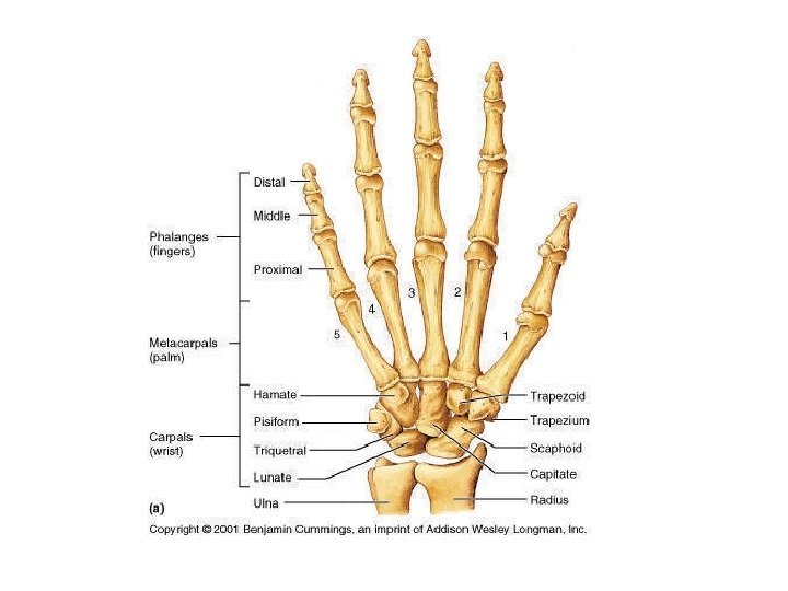
Hand Pelvis Hand Wrist I. Carpals (wrist) A. There are 8 bones in the wrist. The eight bones are known as carpals. B. The carpals are arranged in 2 irregular rows. C. The proximal row contains from lateral to medial the scaphoid, lunate, triquetral and pisiform (smallest carpal). – Remember anatomical position!
D. The distal row contains from lateral to medial the trapezium, trapezoid, capitate, and hamate (large hook). E. Carpal bones are held tightly together by ligaments, their range of motion is limited. F. The scaphoid and the lunate articulate with the distal end of the radius.
II. Hand – The bones of the hand are divided into 2 groups: the metacarpals which are the bones of the palm and the phalanges which are the bones of the fingers. A. Metacarpals (numbered, no names) 1. #1 is the metacarpal on the thumb side. 2. #5 is the metacarpal on the little finger side. 3. The base of the metacarpals articulates with the carpals. 4. The head of each metacarpal joins the proximal phalange of each digit. 5. The shaft of each metacarpal is long and slightly curved. 6. The head of the metacarpals are also known as the “knuckles. ”
B. Phalanges – consists of 14 bones 1. Each digit except the thumb has 3 bones: proximal, middle (intermediate), and distal phalange. 2. The thumb has only 2 bones: proximal and distal phalange.
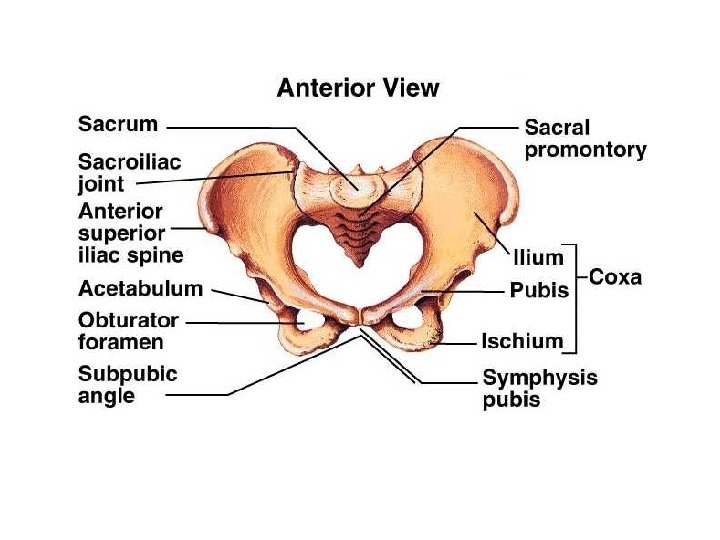
Pelvis (Pelvic Girdle) III. Pelvis A. The pelvic girdle consists of 2 hip bones plus the sacrum and the coccyx. B. The pelvic girdle protects the female reproductive system, some male reproductive organs, nerves and blood vessels to and from the lower extremity, urinary bladder, and the end of the large intestine. C. Each hip bone or os coxae is made up of 3 parts: ilium, ischium, and pubis. D. The femur articulates with the hip bone at the acetabulum and has an acetabular notch. E. The largest foramen in the skeleton called the obturator foramen is found in the pelvic girdle.
IV. Illium A. The ilium has a bowl-shaped body called the iliac fossa. B. Reference points found on the ilium are the iliac crest, anterior superior spine, anterior inferior spine, posterior superior spine, and posterior inferior spine. C. The largest nerve, the sciatic nerve, travels in the greater sciatic notch.
V. Ischium A. The ischium is the most posterior and inferior part of the hip bone. B. You sit on the large tuberosity of the ischium (ischial tuberosity). C. The ischial spine is on the posterior ridge of the ischium. D. Inferior to the ischial spine is the lesser sciatic notch.
VI. Pubis A. The two pubic bones join anteriorly forming the pubic symphysis. B. The obturator foramen is formed from both the ischium and the pubis.
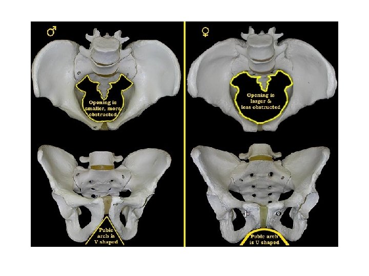
VII. Sex Differences in the Pelvis A. The outlet is bigger in females because a baby’s head needs to pass through. B. In females the pelvis is not as deep. C. In females the sacrum is broader and not so curved. D. The pubic arch is more rounded in females. E. The greater sciatic notch is larger in females. F. The obturator foramen is more triangular in females. G. The acetabulum faces more anteriorly in females. H. The pelvic bones are thinner and lighter in females. I. The spines and ischial tuberosity are more widely spaced in females.