Left Hand Anterior View Carpals proximal Ulna Scaphoid
 Ulna Scaphoid bone Lunate bone Triquetrum bone")


- Slides: 15
Left Hand – Anterior View Carpals (proximal) Ulna Scaphoid bone Lunate bone Triquetrum bone Pisiform bone Radius Hamate bone Capitate bone Trapezoid bone Trapezium bone Carpals (distal) Metacarpals Distal phalanx (of thumb) Phalanges (Digits) Proximal phalanx (of finger) Middle phalanx (of finger) Distal phalanx (of finger)
Right Foot – Superior View Calcaneus Talus Tarsals Cuboid Metatarsals Navicular Medial cuneiform Intermediate cuneiform Lateral cuneiform Proximal phalanx Phalanges (Digits) Middle phalanx Distal phalanx Proximal phalanx (of great toe) Distal phalanx (of great toe)
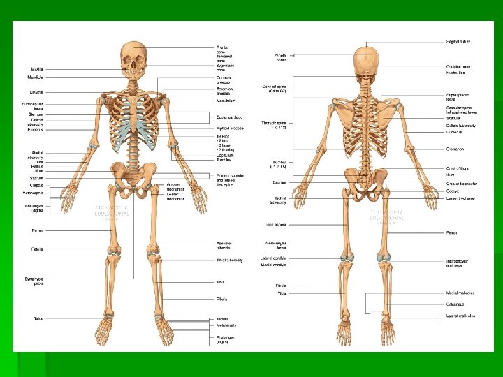
BONE LANDMARKS Bone landmarks are specific locations at which major muscles, ligaments, or other connective tissue attach. The following will familiarize you with basic landmarks and their specific locations on bones.
Surface Markings on Bones Depressions & Openings § Foramen § Fossa
Projections § Process § Condyle § Epicondyle § Head
§ Tuberosity § Tubercle § Trochanter § Crest / Spine
Anatomical Description Landmark Page Reference 2 Foramen i. Foramen magnum § N/A ii. Obturator foramen § Text 25
Anatomical Description Landmark Page Reference 8 Fossa i. Glenoid fossa ii. Supraspinous fossa iii. Infraspinous fossa iv. Subscapular fossa Text 21 § Text 21
Anatomical Description Landmark Page Reference 8 fossa continued v. Olecranon fossa vi. Coranoid fossa vii. Intercondylar fossa vii. Radial Fossa Text 22 § Text 21 § Text 26
Anatomical Description Landmark Page Reference 5 processes i. Coracoid process § Text 21 ii. Acromion process § Text 21 iii. Styloid process § Text 23 iv. Olecranon process § Text 23 v. Coranoid process § Text 23
Anatomical Description Landmark Page Reference 4 condyles i. & ii. Medial condyle § Text 26, 27 iii. & iv. Lateral condyle § Text 26, 27 4 epicondyles i. & ii. Medial epicondyle iii. & iv. Lateral epicondyle Text 22, 26 § Text 22, 26
Anatomical Description Landmark Page Reference 3 heads i. Head of humerus § Text 22 ii. Head of femur § Text 26 iii. Head of fibula § Text 27 Text 22 4 tubercles i. ii. Greater tubercle Lesser tubercle § iii. Infraglenoid tubercle Supraglenoid tubercle § iv. Text 21
Anatomical Description Landmark Page Reference 4 tuberosities i. Deltoid tuberosity § Text 22 ii. Radial tuberosity § Text 23 iii. Ischial tuberosity iv. Tibial tuberosity Text 25 § Text 27 § Text 26 2 trochanters i. ii. Greater trochanter Lesser trochanter
Anatomical Description Landmark Page Reference 9 crests/spines i. Iliac crest Text 25 ii. & iii. Posterior iliac § spines Text 25 iv. Ischial spine § Text 25 v. Anterior crest of § tibia Text 27 vi. Scapular spine Text 21 §