Chapter 49 Sensory and Motor Mechanisms Animal Skeletons


- Slides: 12

• Chapter 49 –Sensory and Motor Mechanisms

Animal Skeletons • Function in support, protection, and movement • Hydrostatic – fluid held under pressure in a closed body compartment (cnidarians, flatworms, nematodes, and annelids) • Exoskeleton – hard encasement deposited on the surface of an animal (molluscs = mantle, calcium carbonate; arthropods = cuticle, chitin) • Endoskeleton – hard supportive elements embedded within the soft tissue

Human Skeleton • 2 main parts: – Axial – skull and vertebral column – Appendicular – limbs, pelvic and pectoral girdles

Human Joints • Ball and socket – where the humerus contacts the shoulder girdle and the femur contacts the pelvic girdle, enable arms and legs to rotate and legs in several planes • Hinge joints – between the humerus and head of the ulna, restrict movement in a single plane • Pivot joints – allows forearm rotation at the elbow and head movement from side to side

Vertebrate Skeletal Muscle • Attached to bone, responsible for movement • Bundles of long fibers running parallel to the length of the muscle • Each fiber is a single cell with multiple nuclei • Myofibrils – smaller fibers that make up a muscle fiber • Myofilaments – make up myofibrils, two types – Thin filaments – actin – Thick filaments - myosin

Vertebrate Skeletal Muscle (cont. ) • Striated – because of regular arrangement of filaments causing light and dark bands • Sarcomere – contractile unit of a muscle • Z line – sarcomere border • M line – center or middle of the sarcomere • I band – only actin, a region of no overlap • A band – actin and myosin protein fibers overlap • H zone – only myosin, center of sarcomere, no overlap

Sliding-Filament Model • Sarcomere length reduced • Myosin = purple, actin = orange • (a) relaxed muscle fiber – the I band H zone are relatively wide • (b) contracting muscle fiber – actin and myosin slide past each other, reducing I band H zone width • (c) fully contracted muscle fiber – H zone and I band are eliminated

Actin/Myosin Interaction • Myosin head is bound to ATP and is in low energy conformation • Myosin head hydrolyzes ATP to ADP and is in high energy conformation • Myosin head binds to actin forming a cross-bridge • Myosin returns to low energy conformation by releasing ADP, slides actin • Binding of new ATP releases myosin from actin and begins a new cycle • Extra energy for contractions stored as creatine phosphate (transfers P to ADP) and glycogen (broken down to make ATP)

Muscle Contraction Regulation • Relaxation: tropomyosin blocks myosin binding sites on actin • Contraction: calcium binds to troponin complex; tropomyosin changes shape, exposing myosin binding sites

Muscle Contraction Regulation (cont. ) • T (transverse) tubules – travel channels in plasma membrane for action potential • Sarcoplasmic reticulum – a specialized endoplasmic reticulum, stores calcium • Stimulated by action potential in a motor neuron, opens calcium channels allowing calcium to enter the cytosol • Ca 2+ then binds to troponin complex triggering a muscle contraction

Review of muscle contraction • ACh released, triggers action potential in muscle fiber • Propagated along membrane and down T tubules • Triggers Ca 2+ release from SR • Ca 2+ bind to troponin, change shape, exposing myosin-binding sites • Myosin cross-bridges attach to actin and detach pulling actin towards center of sarcomere • Ca 2+ is removed by active transport into SR • Tropomyosin blocks myosin binding sites, muscle fiber relaxes

Muscle Contraction Summary • Action potentials and muscle contraction animation • Part 2
 Sensory vs motor
 Grade 5 natural science
 Technology grade 7 term 2 structures
 Pterion
 Sensory
 Cranial nerves sensory and motor
 Nervous
 Dorsal nucleus of vagus
 Motor and sensory nerve
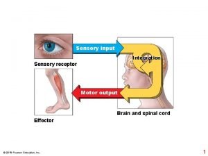
 Incoming sensory impulses and outgoing motor impulses
 How many teeth do adults have on top
 Anatomy quiz
 Dancing skeletons life and death in west africa
