NOTES CH 50 Sensory Motor Mechanisms Sensory Mechanisms


 exteroreceptors: detect stimuli outside the body (heat, light, pressure, chemicals)")


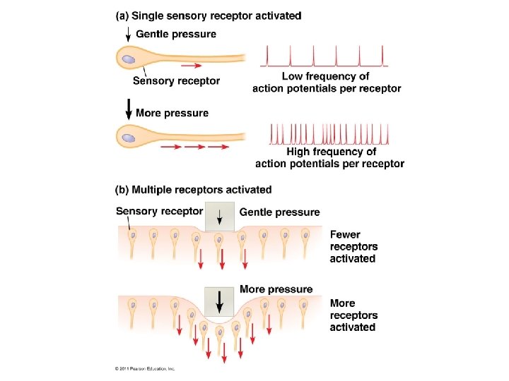
 MECHANORECEPTORS: stimulated by physical deformation (pressure, touch, stretch,")
 3) THERMORECEPTORS: respond to heat or cold; regulate body temp. 4)")


 Rod cells: more sensitive to light (so")


: Vertical pathway: info. passes directly from receptor (i.")


 amplify the")

")


")


 **the")

 extend inward from the")


 PROTEINS INVOLVED: ● TROPONIN and TROPOMYOSIN: together form a complex that covers")
 is released from the")

")


- Slides: 107
NOTES: CH 50 Sensory & Motor Mechanisms!
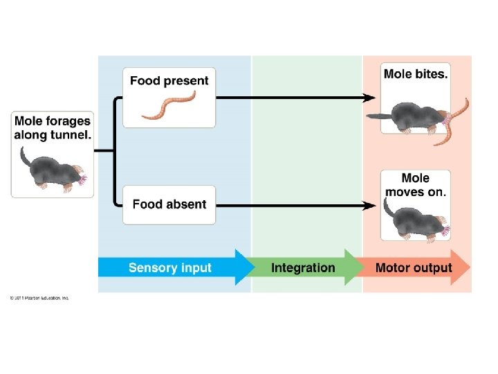
Sensory Mechanisms • Sensations: action potentials that reach the brain via sensory neurons • Perception: interpretation of a sensation *(this occurs in the brain!) “If a tree falls in the woods…. ? ”
Sensory receptors are: 1) exteroreceptors: detect stimuli outside the body (heat, light, pressure, chemicals) 2) interoreceptors: detect stimuli within the body (blood pressure, body position)
The transmission of signals to the nervous system begins with: • SENSORY TRANSDUCTION: -conversion of stimulus energy a change in membrane potential
Sensory transduction is followed by: • AMPLIFICATION: strengthening of stimulus • TRANSMISSION: conduction of impulses to the CNS • INTEGRATION: processing of information (usually in the brain)
5 types of sensory receptors: 1) MECHANORECEPTORS: stimulated by physical deformation (pressure, touch, stretch, motion) 2) PAIN RECEPTORS: stimulated by excess heat, pressure, specific chemicals (like those released by damaged tissues/cells)
Sensory receptors: (continued) 3) THERMORECEPTORS: respond to heat or cold; regulate body temp. 4) CHEMORECEPTORS: detect and transmit info. about solute concentration; involved w/taste & smell; osmoreceptors that regulate kidneys &rate of urine production 5) ELECTROMAGNETIC RECEPTORS: include photoreceptors (detect light); used in some animals for migration (use magnetic field of earth)
CHEMORECEPTORS in an insect!
CHEMORECEPTORS:
ELECTROMAGNETIC RECEPTORS:
ELECTROMAGNETIC RECEPTORS:
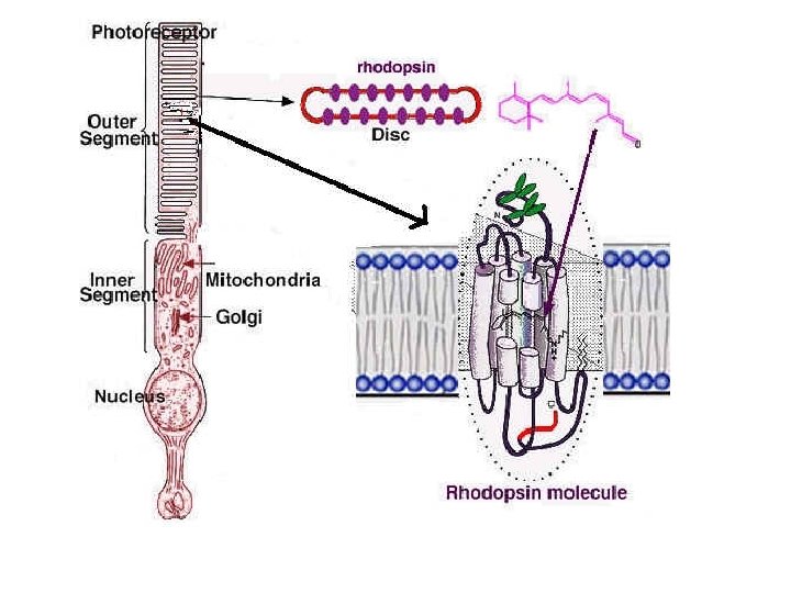
PHOTORECEPTORS: • PHOTORECEPTORS: cells that contain pigment molecules that absorb light; transduce the light stimulus into an electrical signal (by changing the membrane potential of the cell)
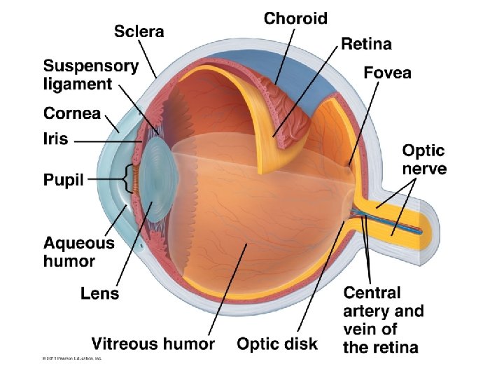
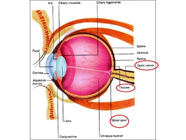
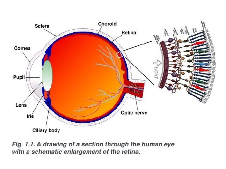
Vertebrate EYE: see fig. 50. 17 Structure Function Sclera tough outer layer of connective tissue Choroid thin inner pigmented layer Cornea anterior, transparent area of sclera; Iris anterior choroid; regulates amt. of light Pupil hole in center of iris Conjunctiva Mucous membrane covering the sclera; keeps the eye moist allows light to enter eye entering pupil; gives eye its color
Vertebrate EYE, continued Retina innermost layer of eyeball; contains Lens transparent, protein disc that Ciliary body produces aqueous humor Aqueous humor fills cavity between lens and cornea; fxns as liquid lens; clear & watery Vitreous humor fills cavity behind lens; fxns as liquid lens; jellylike Optic nerve carries visual sensory info to brain photoreceptor cells focuses image on retina
Vertebrate EYE, continued Fovea Center of the visual field; near the optic nerve; densely packed with cone cells Blind spot Where the optic nerve exits the eye – no photoreceptors here
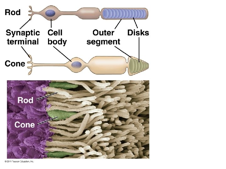
Two types of photoreceptors on retina: 1) Rod cells: more sensitive to light (so they help us to see in dim light); do not distinguish colors; allow us to see at night; more numerous in nocturnal animals (approx. 125 million; make up 70% of all sensory receptors in the body!) 2) Cone cells: do not function at night; can distinguish colors in daylight; approx. 6 million
*Rods and cones contain visual pigments which consist of light-absorbing pigment molecules (retinal…derived from vitamin A) and a membrane protein (opsin). Retinal Opsin
RHODOPSIN: ● the visual pigment of ROD CELLS ● has 2 alternating forms depending on the conditions: DARK or LIGHT
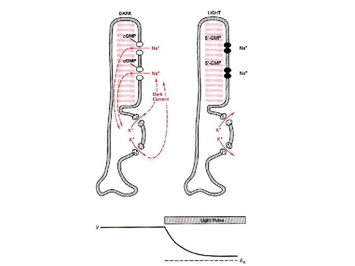
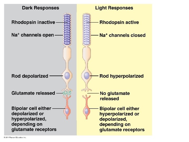
IN THE DARK… ● rhodopsin is INACTIVE. ● rod cells are highly permeable to sodium and are therefore in a DEPOLARIZED state ● rod cells in this INACTIVE (dark) state are releasing neurotransmitter molecules that INHIBIT the firing of postsynaptic cells in the retina. ● so in the dark, no message is sent from the rod cells to the visual centers of the brain
IN THE LIGHT… ● rhodopsin absorbs light, and breaks apart, as its retinal component changes shape; opsin is now ACTIVE; ● this triggers a chain of metabolic events (signaltransduction pathway!) that makes the rod cell membrane less permeable to sodium and therefore hyperpolarizes the rod cell membrane; ● the rod cell synaptic terminals stop releasing neurotransmitter (which was inhibiting the postsynaptic cell);
IN THE LIGHT… ● thus, a decrease in chemical signal to the cells with which the photoreceptors synapse serves as the message that rods have been stimulated by light…these postsynaptic neurons, now freed from inhibition, can develop action potentials which are transmitted to the brain for processing
Photoisomerization of rhodopsin:
● over time, very bright light keeps the rhodopsin “bleached” (most of the rhodopsin decomposes into retinal and opsin) and rod cells eventually become unresponsive cone cells take over; ● in the dark, enzymes convert the retinal back to its original form (rhodopsin) and the rod cells can once again respond to faint light (e. g. walking from a bright environment into a dark room or movie theatre…)
CONES and COLOR VISION • there are 3 subclasses of cone cells, each with a different opsin protein • each photopsin is best at absorbing a specific wavelength (color) of light • the 3 subclasses are: red green blue
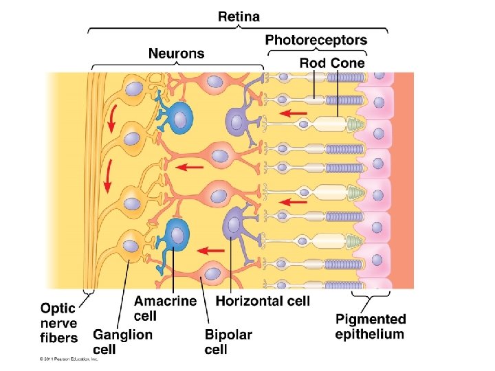
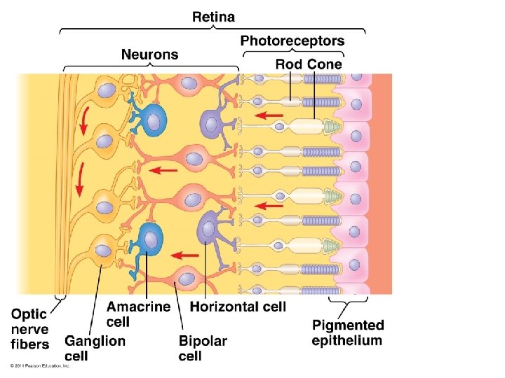
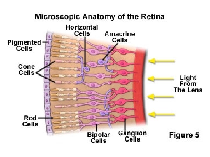
Integration of visual information: • begins in the retina • rod and cone cells synapse with: BIPOLAR CELLS, which synapse with: GANGLION CELLS • HORIZONTAL CELLS and AMACRINE CELLS also involved
Integration of visual info (cont. ): Vertical pathway: info. passes directly from receptor (i. e. the rod or cone cell) to bipolar to ganglion cells Lateral pathway: info. passes from rod/cone to horizontal/amacrine cells and spreads out over several bipolar or ganglion cells *(nearby cells are stimulated; distant receptor & bipolar cells are inhibited “lateral inhibition”…sharpens the edges of objects)
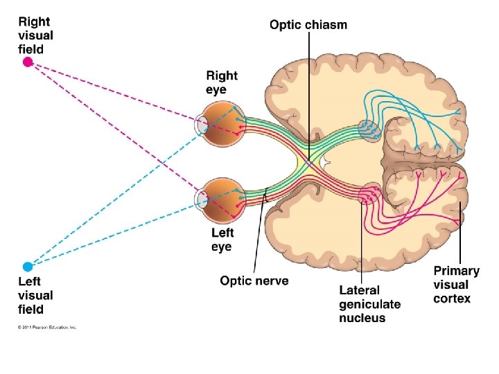
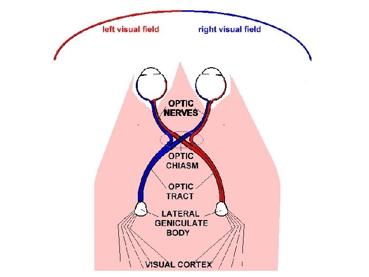
• Finally, the info. is transmitted along the optic nerves formed by axons of ganglion cells; • optic nerves from the 2 eyes meet at the OPTIC CHIASM;
• they pass through the thalamus; • they continue back to the primary visual cortex in the occipital lobe of the cerebrum (integration occurs here and in other cortex areas)
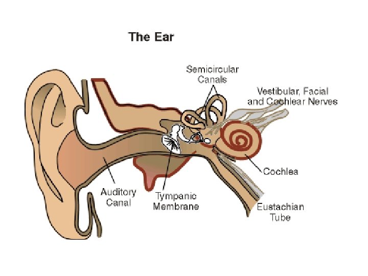
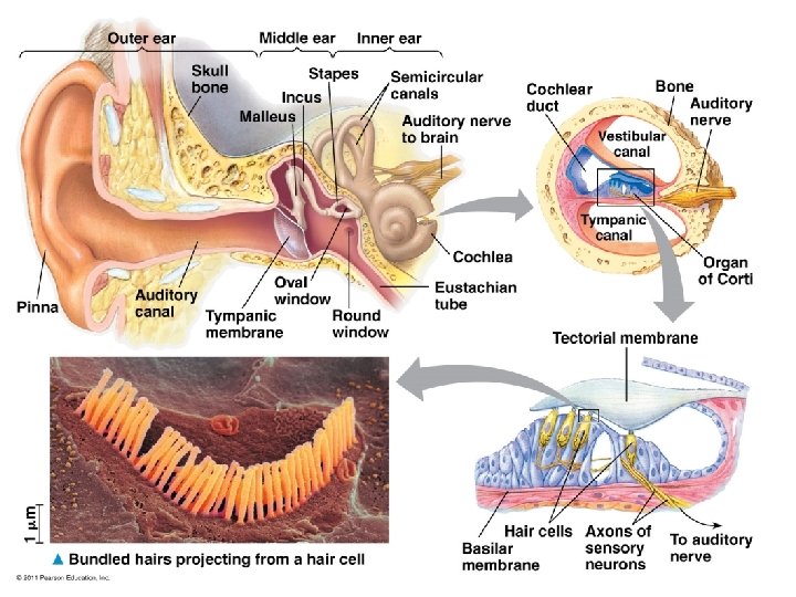
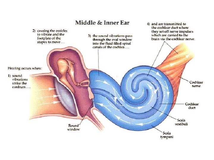
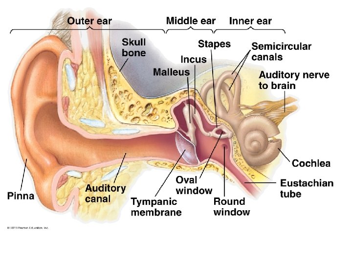
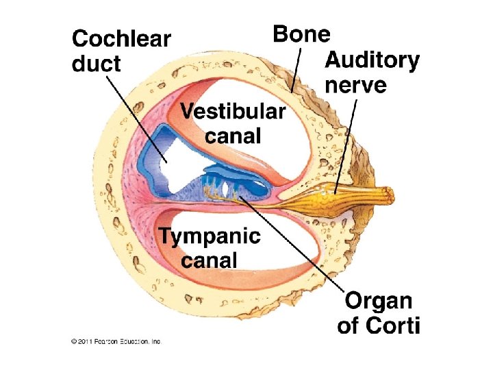
HEARING AND EQULIBRIUM: • vibrating objects create percussion waves in air; • these waves cause the tympanic membrane (EARDRUM) to vibrate w/same freq. ;
• the 3 bones of the middle ear (malleus, incus, stapes) amplify the sound and transmit the mechanical movements to the oval window (membrane covering cochlea in the inner ear); • vibrations of oval window produce pressure waves in the fluid w/i the cochlea; • mechanoreceptors in the cochlea convert the energy of the vibrating fluid into action potentials (which travel on the auditory pathway to the cerebral cortex)
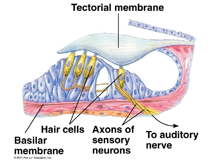
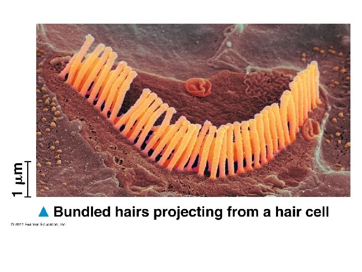
The receptor cells – hair cells – are part of the organ of Corti; as hair cells are physically moved by the sound waves (transmitted through the cochlear fluid), they brush against the tectorial membrane; this causes them to depololarize and release neurotransmitter, thereby triggering an action potential in a sensory neuron!
*(the middle ear also contains the Eustachian tube which connects with the pharynx)
*Equilibrium and balance are affected by the semicircular canals in the inner ear
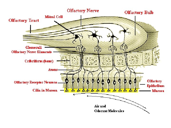
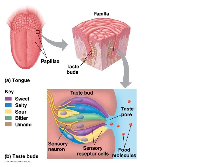
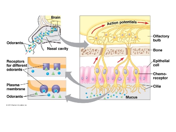
CHEMORECEPTION: Taste and Smell • chemoreceptors in taste buds or nasal cavity are triggered when specific molecules bind and trigger action potentials **4 basic taste perceptions: sweet, sour, salty, bitter • taste and smell interact; if the olfactory system is blocked (e. g. you have a cold) the perception of taste is sharply reduced
**Example of chemoreceptors…solutes in air dissolve in mucus; bind to chemoreceptors; result in the opening of ion channels; sensory neuron is depolarized; action potential goes to the brain via the olfactory nerve…you perceive a SMELL!
(He thought it was funny)
CH 50, continued… Muscles & Movement
● Movement is necessary to: -catch food -escape predators -locate mates
● Skeletons are essential to movement: -they provide a firm attachment against which muscles can work during movement
The muscular system consists of three types of muscle tissue: ● Skeletal ● Smooth ● Cardiac
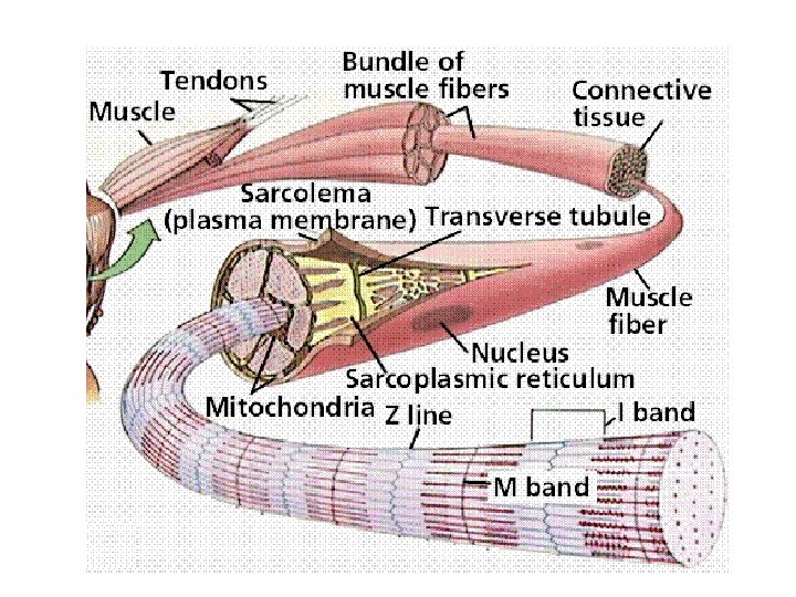
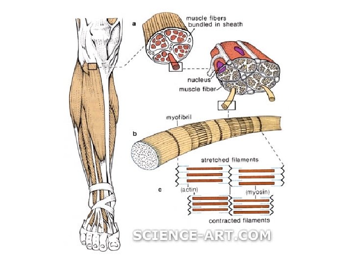
STRUCTURE OF A SKELETAL MUSCLE: *Individual muscles are the organs of the muscular system. They contain skeletal muscle tissue, nervous tissue, blood, and connective tissues.
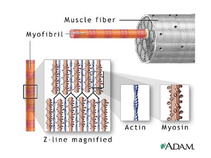
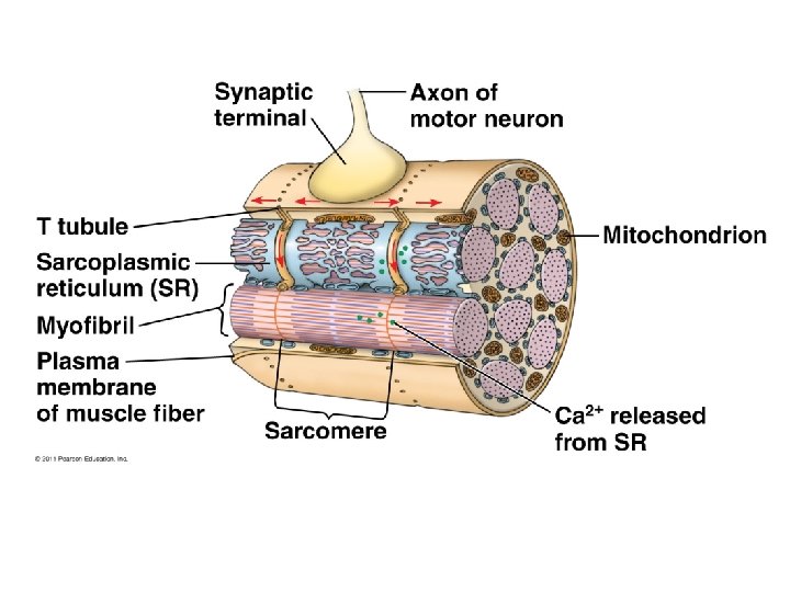
Skeletal Muscle Fibers: each muscle fiber is a single muscle cell, which is the unit of contraction just beneath the cell membrane the cytoplasm (SARCOPLASM) contains: * many small, oval nuclei * mitochondria * SARCOPLASMIC RETICULUM (a modified endoplasmic reticulum) * MYOFIBRILS (fibers of the proteins ACTIN and MYOSIN)
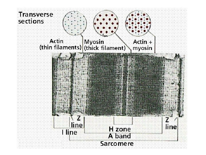
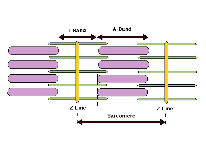
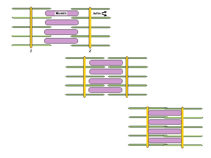
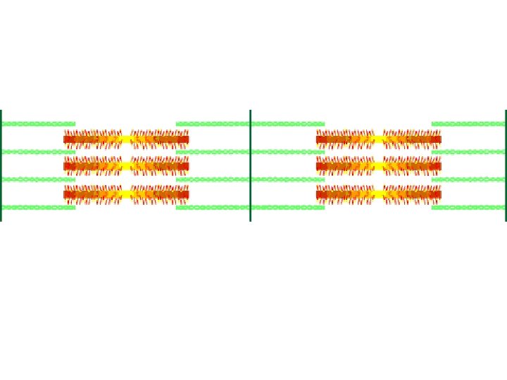
Skeletal Muscle Fibers: **the organization of actin and myosin filaments produces STRIATIONS (bands) **the thick (myosin) and thin (actin) filaments are organized into structural units called SARCOMERES
SARCOMERE: unit of organization of skeletal muscle ● Z lines: borders of sarcomere ● I bands: area near the edge; contains only thin filaments ● A bands: regions where thick and thin filaments overlap ● H zones: areas in center of A bands containing only thick filaments
Also part of a muscle fiber… **TRANSVERSE TUBULES (T tubules) extend inward from the cell membrane and associate with the SARCOPLASMIC RETICULUM (whose membranes surround each myofibril)
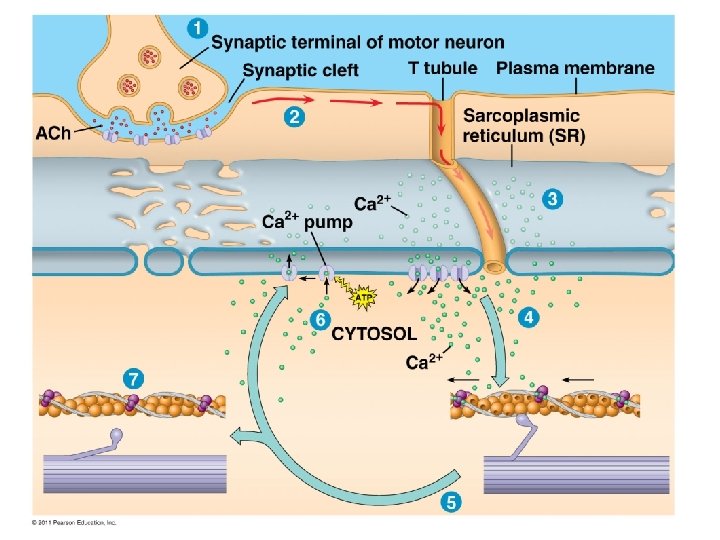
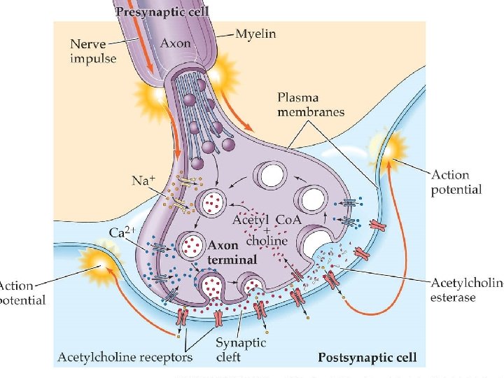
Neuromuscular Junction: MOTOR NEURONS stimulate muscle fibers to contract in response to a nerve impulse, the end of a motor neuron axon secretes a NEUROTRANSMITTER, which stimulates the muscle fiber to contract
Neuromuscular Junction: one MOTOR NEURON and the MUSCLE FIBERS associated with it constitute a MOTOR UNIT all muscle fibers of a motor unit contract together!
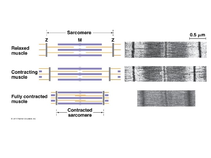
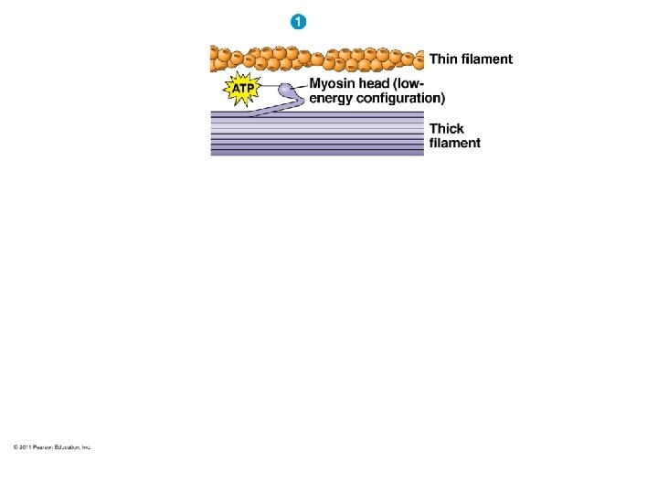
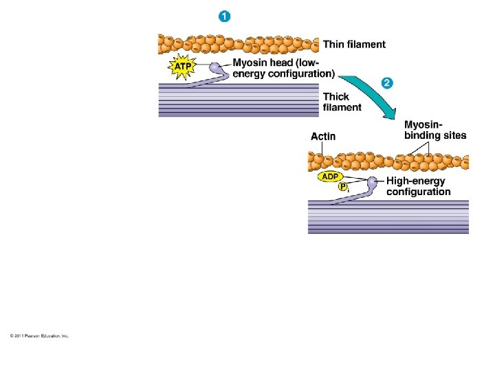
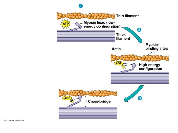
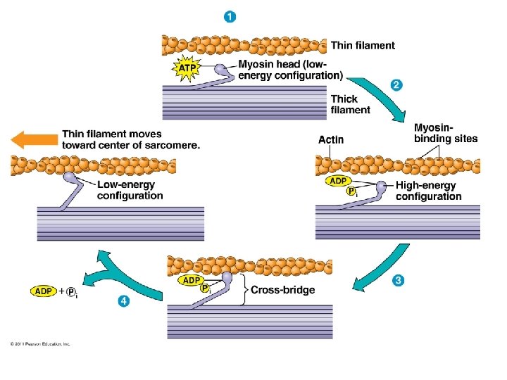
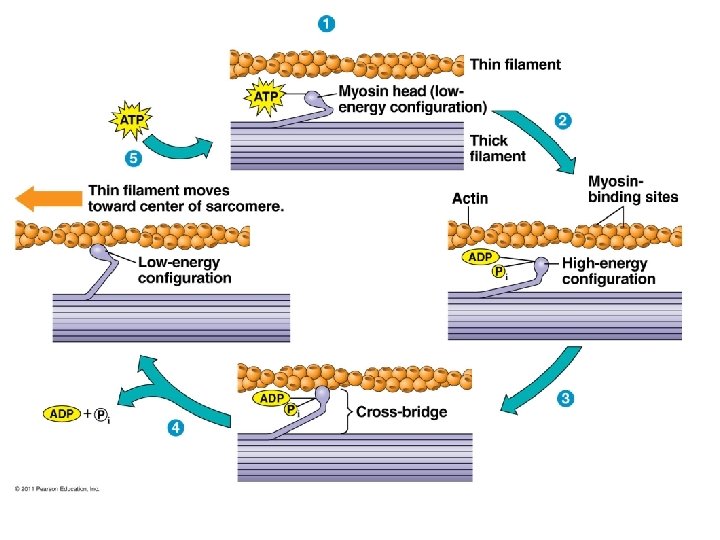
SKELETAL MUSCLE CONTRACTION *Muscle fiber contraction results from a sliding movement of actin and myosin filaments. (known as the SLIDING FILAMENT MODEL in which individual sarcomeres shorten)
Role of MYOSIN and ACTIN: cross-bridges of myosin filaments form linkages with actin filaments the reaction between actin and myosin filaments generates the force of contraction
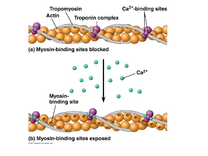
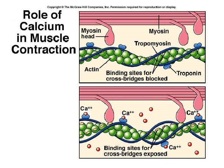
OTHER (REGULATORY) PROTEINS INVOLVED: ● TROPONIN and TROPOMYOSIN: together form a complex that covers the myosin-binding sites on actin; by covering these binding sites, myosin cannot bind to actin and a contraction cannot occur.
Stimulus for and Steps of…a CONTRACTION: > ACETYLCHOLINE (a neurotransmitter) is released from the distal end of a motor neuron axon and stimulates a skeletal muscle fiber > acetylcholine causes the muscle fiber to conduct an impulse (membrane potential) over the surface of the fiber that reaches deep within the fiber through the TRANSVERSE TUBULES > a muscle impulse signals the sarcoplasmic reticulum to release CALCIUM IONS
Steps of a Muscle Contraction… > calcium ions bind to troponin protein & tropomyosin is pulled aside, uncovering the myosin-binding sites on actin > linkages form between actin and myosin > the myosin cross-bridges pull on actin filaments, shortening the fiber ****energy for the sliding filament model comes from ATP!!!
The end of a contraction… > the muscle fiber relaxes (and the contraction ends) when cross-bridges release from actin and when calcium ions are actively transported back into the sarcoplasmic reticulum (without calcium present, the troponin-tropomyosin complex re-covers the myosin-binding sites on actin) > acetylcholine is broken down by the enzyme ACETYLCHOLINESTERASE
Energy Sources for Muscles: ● ATP supplies the energy for muscle fiber contraction ● for sustained muscle contractions, a molecule called creatine phosphate is used to make more ATP
Oxygen Supply and Cellular Respiration ● aerobic respiration requires oxygen ● red blood cells carry oxygen to body cells (oxygen binds to HEMOGLOBIN in the RBCs) ● MYOGLOBIN in muscle cells temporarily stores oxygen
Oxygen Debt ● during rest or moderate exercise, muscles receive enough oxygen to respire aerobically ● during strenuous exercise, oxygen deficiency may cause LACTIC ACID to accumulate ● OXYGEN DEBT is the amount of oxygen required to convert accumulated lactic acid to glucose and to restore supplies of ATP **the metabolic capacity of a muscle may change with training!
Muscle Fatigue: ● a fatigued muscle loses its ability to contract ● muscle fatigue is usually due to accumulated lactic acid
Heat Production **muscle action is an important source of body heat!